. 2007 Jan;170(1):176-87.
doi: 10.2353/ajpath.2007.051212.

Identification and functional characterization of the hepatic stellate cell CD38 cell surface molecule

Affiliations

Identification and functional characterization of the hepatic stellate cell CD38 cell surface molecule

Sandra March et al. Am J Pathol. 2007 Jan.

Abstract

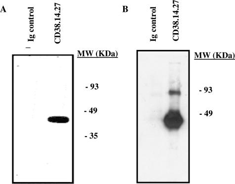
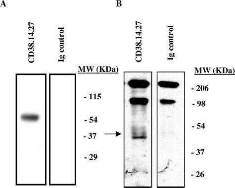
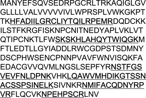
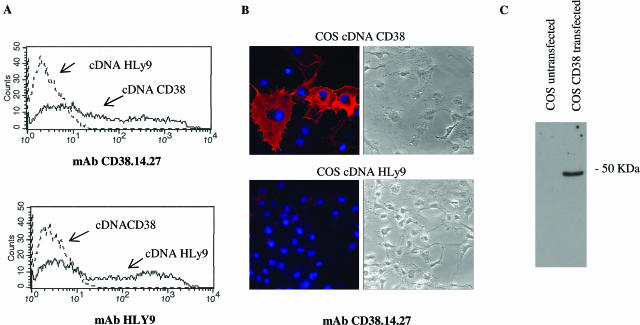
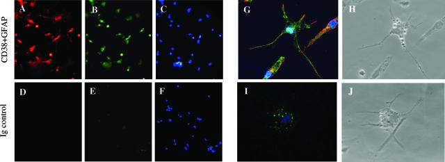
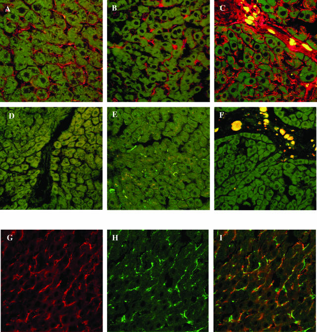
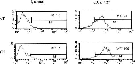
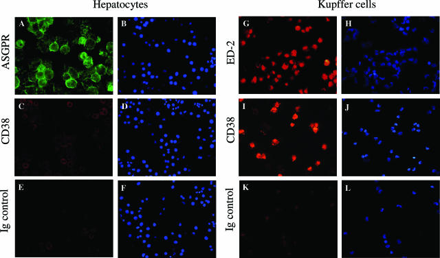
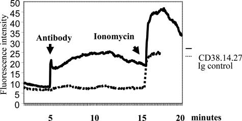
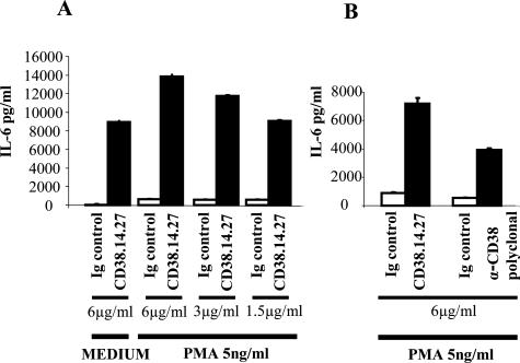
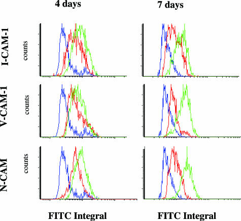
The activation of hepatic stellate cells (HSCs) is a critical event in hepatic fibrosis, because these cells are the main producers of extracellular matrix proteins in the liver and contribute to the modulation of inflammatory responses via the secretion of several cytokines and the expression of adhesion molecules. The goal of the present study was to characterize cell surface proteins that regulate HSC activation. To this end, a panel of monoclonal antibodies (mAbs) was generated. mAb 14.27 recognized a protein of 45 kd that was highly expressed on HSCs. Affinity purification of this protein followed by sequencing revealed that protein to be CD38. We subsequently demonstrated that CD38 was constitutively expressed by HSCs and that its expression increased after in vitro and in vivo activation. mAb 14.27 induced an increase in cytosolic Ca2+ levels in HSCs, showing that it functions as an agonistic antibody. Moreover, the effects mediated by the CD38 mAb included induction of the proinflammatory cytokine interleukin-6 and up-regulation of the adhesion molecules intercellular adhesion molecule-1, vascular cell adhesion molecule-1, and neural cell adhesion molecule. Collectively, our data suggest that CD38 can act as a regulator of HSC activation and effector functions.


Figures

Figure 1
Figure 1
Immunoprecipitation of the cell surface protein recognized by mAb 14.27. Detergent lysates of surface-labeled HSCs were immunoprecipitated with 14.27 mAb or with an irrelevant mAb. The immunoprecipitated material was analyzed under nonreducing conditions on a 12% SDS-polyacrylamide gel. Two independent immunoprecipitations are shown at two different exposure times (A, short exposure time; B, long exposure time). Molecular masses (in kilodaltons) were determined by the migration of a protein standard.
Figure 2
Figure 2
Western blot analysis of the protein recognized by mAb 14.27. Detergent lysates of HSCs (25 μg) (A) or liver tissue (100 μg) (B) were analyzed by Western blotting (12% SDS-polyacrylamide gel) using an anti-CD38 mAb (14.27). Molecular masses (in kilodaltons) were determined by the migration of a protein standard.
Figure 3
Figure 3
Amino acid sequence of rat CD38. CD38 was identified by peptide-mass fingerprinting using MALDI-TOF mass spectroscopy. The tryptic peptides obtained from the 45-kd band matched the rat CD38 amino acid sequence (UniProtKB accession no. Q64244) and covered 33% of the protein. Darks lines indicate amino acids predicted by mass spectrometry.
Figure 4
Figure 4
Reactivity of 14.27 mAb with CD38 cDNA-transfected COS cells. A: COS cells transfected with rat CD38 cDNA (solid lines) or human HLy9 cDNA (dotted lines) were stained with 14.27 and analyzed by flow cytometry (top). As a positive control, COS cells transfected with rat CD38 cDNA (dotted lines) or human HLy9 cDNA (solid lines) were stained with HLY9 mAb (bottom). The fluorescence intensity is shown over a 3-decade log scale. B: COS cells transfected cells with rat CD38 cDNA (top) or human HLy9 cDNA (bottom) were washed with PBS, fixed with paraformaldehyde, and incubated with 14.27 mAb. Cells were washed and incubated with a Cy-3-conjugated secondary antibody (red). Nuclei were stained with Hoechst reagent (blue). Fluorescence images were acquired using a confocal spectral microscope with an original magnification of ×63. C: Detergent lysates of COS untransfected and COS CD38-transfected cells were analyzed by Western blotting (12% SDS-polyacrylamide gel) using anti-CD38 mAb (14.27). Molecular mass (in kilodaltons) was determined by the migration of a protein standard.
Figure 5
Figure 5
Expression of CD38 and the HSC-specific marker GFAP on cultured HSCs. HSCs were maintained for 3 days in culture. Cells were washed with PBS, fixed with paraformaldehyde, and permeabilized. Double immunostaining was performed. Cells were incubated with CD38.14.27 mAb and a rabbit anti-GFAP polyclonal antibody (AC, G, and H, top) or with Ig controls (DF, I, and J, bottom). Cells were washed and incubated with an anti-mouse Cy-3-conjugated secondary antibody (red) and an anti-rabbit Cy-2-conjugated secondary antibody (green). I: Vitamin A autofluorescent droplets were visualized in the negative control. C and F: Nuclei were stained with Hoechst reagent (blue). H and J: Phase contrast microscopic view. Fluorescence images were acquired using a fluorescence microscope with an original magnification of ×40 (AF) or using a confocal spectral microscope with an original magnification of ×63 (GJ).
Figure 6
Figure 6
Immunolocalization of CD38 in rat liver sections. AF: Liver sections of control rats (A and D), rats pretreated with LPS (550 μg/rat) (B and E) and cirrhotic rats with an advanced stage of fibrosis displaying prominent scars (C and D) were stained with an antibody against CD38 (CD38.14.27) (AC) or with an Ig control (DF). Liver sections were washed and incubated with an anti-mouse Cy-3-conjugated secondary antibody (red). Hepatocyte autofluorescence from hepatocytes is seen in green. GI: Double-immunostaining was performed. Liver sections of control rats were incubated with mAb CD38.14.27 (G) and a rabbit anti-GFAP polyclonal antibody (H). Merged image is seen in I. Liver sections were washed and incubated with an anti-mouse Cy-3-conjugated secondary antibody (red) and an anti-rabbit Cy-2-conjugated secondary antibody (green).
Figure 7
Figure 7
Expression of CD38 on activated HSCs. HSCs were freshly isolated from control (top) and cirrhotic (bottom) rats. CD38+ cells were gated, and the intensity of CD38 expression was assessed by flow cytometry (FACS) using CD38.14.27 mAb. Fluorescence intensity is shown over a 3-decade log scale.
Figure 8
Figure 8
CD38 expression in endothelial cells in the liver. Rat liver sections from central venule (A) and portal areas (B) were stained with the antibody CD38.14.27 (A and B), RECA (pan-endothelial marker) (C) or with an Ig control (D). Sections were washed and incubated with an anti-mouse Cy-3-conjugated secondary antibody (red). Hepatocyte autofluorescence from hepatocytes and vessels shown in green. EM: Sinusoidal endothelial cells were isolated from rat livers and cultured for 2 days. Cells were washed with PBS, fixed with paraformaldehyde, and single immunostaining was performed. Cells were incubated with CD38.14.27 mAb (E, H, and K), RECA (F, I, and L), or Ig controls (G, J, and M). H and J: Cells were washed and incubated with an anti-mouse Cy-3-conjugated secondary antibody (red). Corresponding phase-contrast images are shown (EG) and corresponding nuclei are seen by Hoechst staining (KM). Fluorescence images were acquired using a confocal spectral microscope with an original magnification of ×40 (AD) or using a fluorescence microscope with and original magnification of ×20 (EM).
Figure 9
Figure 9
CD38 expression in hepatocytes and Kupffer cells. AF: Hepatocytes. Freshly hepatocytes were isolated and cytospun followed by single immunostaining. Hepatocytes were incubated with rabbit ASGPR polyclonal antibody (A and B), CD38.14.27 mAb (C and D), and Ig control (E and F). Cells were then washed and incubated with an anti-rabbit Cy-2-conjugated secondary antibody (green) (A and B) or an anti-mouse Cy-3-conjugated secondary antibody (red) (CF). Corresponding nuclei are shown by Hoechst staining (blue). Ig control incubated with anti-rabbit Cy-2-conjugated secondary antibody (data not shown). GL: Kupffer cells. Fresh Kupffer cells were isolated, and single immunostaining was performed. Kupffer cells were incubated with ED-2 mAb (G and H), mAb CD38.14.27 (I and J), and Ig control (K and L). G and L: Cells were then washed and incubated with an anti-mouse Cy-3-conjugated secondary antibody (red). Corresponding nuclei are shown by Hoechst staining (blue). Fluorescence images were acquired using a fluorescence microscope with an original magnification of ×40.
Figure 10
Figure 10
Ca2+ mobilization in HSCs by CD38 ligation. HSCs cultured for 6 days were loaded with the fluorescent indicator Fluo-4/AM. Cells were incubated with mAb CD38.14.27 (6 μg/ml) and immediately analyzed using an inverted confocal microscope. The image was analyzed using software Image Processing Leica Confocal Software. The same experiment was performed with an isotype-matched mAb (IgG2b).
Figure 11
Figure 11
Secretion of IL-6 by HSCs followed by CD38 ligation. A: HSCs were incubated in the presence or absence of PMA (5 ng/ml) and with mAb CD38.14.27 at different concentrations (6, 3, and 1.5 μg/ml). The same concentrations of an isotype-matched mAb (IgG2b) were used as controls. B: HSCs were incubated in the presence of PMA (5 ng/ml) and 6 μg/ml mAb CD38.14.27 or a polyclonal anti-CD38 antibody. The same concentrations of an isotype-matched antibody (IgG2b or goat IgG) were used as controls.
Figure 12
Figure 12
Expression of ICAM-1, VCAM-1, and NCAM in HSCs followed by CD38 ligation. HSCs cultured for 3 or 6 days were incubated for 12 hours with mAb CD38.14.27 or with an isotype-matched mAb (IgG2b) as a control. Expression of these adhesion molecules was assessed by LSC cytometry using the primary and secondary antibodies described in Materials and Methods. Lines in the histogram: negative control (blue), isotype-matched control activation (red), and CD38 ligation (green).

References

    1. Geerts A. History, heterogeneity, developmental biology, and functions of quiescent hepatic stellate cells. Semin Liver Dis. 2001;21:311–335. - PubMed
    1. Friedman SL. Molecular regulation of hepatic fibrosis, an integrated cellular response to tissue injury. J Biol Chem. 2000;275:2247–2250. - PubMed
    1. Albanis E, Friedman SL. Hepatic fibrosis: pathogenesis and principles of therapy. Clin Liver Dis. 2001;5:315–334, v-vi. - PubMed
    1. Friedman SL. Seminars in medicine of the Beth Israel Hospital, Boston. The cellular basis of hepatic fibrosis: mechanisms and treatment strategies. N Engl J Med. 1993;328:1828–1835. - PubMed
    1. Bataller R, North KE, Brenner DA. Genetic polymorphisms and the progression of liver fibrosis: a critical appraisal. Hepatology. 2003;37:493–503. - PubMed

Publication types

MeSH terms