. 2007 Jan;170(1):334-46.
doi: 10.2353/ajpath.2007.060447.

Tumor cell transendothelial passage in the absorbing lymphatic vessel of transgenic adenocarcinoma mouse prostate

Affiliations

Tumor cell transendothelial passage in the absorbing lymphatic vessel of transgenic adenocarcinoma mouse prostate

Giacomo Azzali. Am J Pathol. 2007 Jan.

Abstract

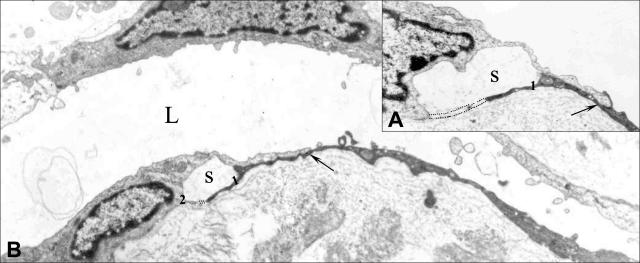
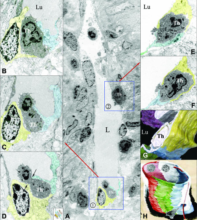
The distribution and fine structure of the tumor-associated absorbing lymphatic vessel in the tumor mass of prostate adenocarcinoma and of seminal vesicle metastasis in transgenic mice was studied for the purpose of understanding the modality of tumor cell transendothelial passage from the extravasal matrix into the lymphatic vessel. In the tumor mass, two main cell populations were identified: stromal tumor cells and the invasive phenotype tumor (IPT) cells, having characteristics such as a highly electron-dense matrix rich in small granules lacking a dense core and massed nuclear chromatin, which is positive to immunostaining with anti-SV40 large T antigen antibody. Based on the ultrastructural pictures of different moments of the IPT cell transendothelial passage by ultrathin serial sections of the tumor-associated absorbing lymphatic vessel, the manner of its transendothelial passage through the intraendothelial channel, without involving intercellular contacts, was demonstrated. The presence of IPT cells in the parenchyma of satellite lymph node highlights its significant role in metastatic diffusion. The intraendothelial channel is the reply to the lack of knowledge regarding the intravasation of the tumor cell into the lymphatic circulation. The lymphatic endothelium would organize this channel on the basis of tumor cell-endothelial cell-extravasal matrix molecular interactions, which are as yet unidentified.


Figures

Figure 1
Figure 1
Morphological aspects useful to compare shape and sizes of prostate and seminal vesicles of wild-type and transgenic mice. A: Prostate (P) and seminal vesicles of 4-month-old wild-type mouse. B: Prostate (P) and hypertrophic seminal vesicles with metastatic area (frame) of 6-month-old TRAMP mouse.
Figure 2
Figure 2
Histological, histochemical, and ultrastructural aspects of prostate tumor mass of 6-month-old TRAMP mouse. A and B: Peripheral part of the tumor mass with peritumoral fibromuscular stroma (P) containing a tumor-associated lymphatic vessel TAAL (L) surrounded by tumor cells (circles), cytoplasms of which have a strong dye-affinity for toluidine blue. C and D: IPT cells (Th) characterized by filopods, undulopods, and by peculiar ultrastructural aspects of the cytoplasmatic matrix that is rich in dense granules, nucleus of which (E) is immunohistochemically positive to SV40 large T-antigen protein (colored dark brown, arrows). Coloration is toluidine blue, original magnification: ×1920 (A); ×3100 (B); ×17,500 (C); 1/4 original magnification, ×26,500; 1/2 original magnification (D). Mayer’s hematoxylin, original magnification, ×920 (E).
Figure 3
Figure 3
Ultrastructural aspects of TAAL vessel and its relationship with stromal tumor cells and IPT cells. A: TAAL vessel (L) of the prostate tumor mass, under oblique direction of the section plane (small inset at bottom), formed of two endothelial cells whose nonnuclear cytoplasmatic expansions are joined by overlapping contacts (arrows) fixed by tight and gap junctions. T, stromal tumor cell; Th, IPT cell close to the lymphatic endothelial wall. B: TAAL vessel (L) of the metastatic mass of the seminal vesicles, characterized by the peculiar alternating disposition of endothelial cells with an electron-dense matrix and cells with a light matrix, whose ultrastructural characteristics (rectangle) can be better seen in C. In the same figure, the behavior of the cytoplasmatic expansions of adjacent cells in circumscribing an interstitial space (S) can be observed. Arrows: overlapping contacts between interendothelial cells fixed by tight and gap junctions; Lu, lymphatic vessel lumen. D: Lymphatic endothelial cell with electron-dense cytoplasmatic matrix expansion having many vacuoles (arrows), some of which contain synaptic-type microvesicles (E). N, nucleus; Lu, lymphatic vessel lumen. Original magnifications: ×12,960 (A); ×14,500 (B); ×26,500 (C); ×14,000 (D); ×41,500 (E).
Figure 4
Figure 4
Metastatic tumor mass of the seminal vesicle with TAAL vessel in almost sagittal section plane surrounded by stromal tumor cell (T) and with an IPT cell embedded in the endothelial wall. A: In frame 1, the IPT cell (Th) appears embedded in the endothelial wall, whereas in frame 2 the IPT cell is inside the lumen of the vessel. B and C: The ultrastructural aspects can be observed. The IPT cell of frame 1 in C is inserted between the nonnuclear cytoplasmatic expansions 1 and 2 of adjacent endothelial cells (colored yellow and light blue). Arrow, interendothelial contact fixed by tight and gap junctions. Original magnifications: ×3100 (A); ×19,000 (B); ×21,000 (C).
Figure 5
Figure 5
Ultrastructural pictures of a TAAL vessel (L) chosen from 96 ultrathin serial sections to demonstrate the manner of intraendothelial channel formation. A and B: The secondary extension (marked 1) of the nonnuclear cytoplasmatic expansions of an endothelial cell and its elongation (dashed line) in the interstitial matrix circumscribing a space (S), ending in the secondary extension of the adjacent endothelial cell (marked 2). This behavior anticipates the formation of an intraendothelial channel. Arrows, overlapping interendothelial contacts fixed by tight and gap junctions. Original magnifications: ×26,500 (A); ×12,500 (B).
Figure 6
Figure 6
Metastatic tumor mass of the seminal vesicles with TAAL vessel (L) surrounded by stromal tumor cells (T) and by two IPT cells (Th), one of which is attached to the endothelial abluminal wall and the other shown in a moment of its transendothelial passage through an intraendothelial channel. A, B, and C demonstrate, in an ultrathin serial section at a different level, the same Th cell (frame) in its passage between the nonnuclear cytoplasmic expansions of adjacent endothelial cells (arrow and arrowhead); this morphological aspect results from the sagittal direction of the section plane of the intraendothelial channel (bottom right inset). Original magnifications: ×4700 (A); × 11,200 (B); ×26,500 (C).
Figure 7
Figure 7
TAAL vessel (L) under sagittal direction of the section plane to show two IPT cells embedded in the endothelial wall. A: Frames 1 and 2 show the passage of IPT cell through the endothelial wall at different moments. B, C, and D show ultrastructural pictures chosen from 220 ultrathin serial sections of the transendothelial passage of the IPT cell (Th) in frame 1 of A into the lymphatic vessel lumen (Lu) through the intraendothelial channel (see Results for details). Inset in D denotes sagittal direction of the section plane. Arrows show the cell body restriction. E and F show serial sections of the IPT cell in frame 2 of A in the initial phase of its transendothelial passage from the extravasal matrix into the lymphatic vessel lumen (Lu), as documented also by the three-dimensional reconstruction in G. H: Schematic drawing of a TAAL vessel to illustrate, through synthesis of the various ultrastructural pictures of cytoplasmatic expansions of adjacent endothelial cells, how the intraendothelial channel is formed without the participation of intercellular junctions. The dashed line and the arrow underlie the abluminal-luminal direction of the cancer cell transendothelial passage. Original magnifications: ×3100 (A); ×14,500 (B); ×12,000 (C, D); ×8100 (E); ×9300 (F).
Figure 8
Figure 8
Ultrastructural pictures of parailiac lymph node parenchyma of 6-month-old TRAMP mouse demonstrating the presence of IPT cells. A and B: IPT cells (Th, small circles) distributed between the endothelium (en) that covers the cells of the cortical area, and the marginal sinus (S). L, small and medium lymphocytes; m, macrophage with dense bodies (phagosomes, lysosomes); r, reticular phagocytic cell; e, eosinophil leukocyte; arrows, connective capsule. CE: IPT cells (Th) distributed between cells forming the cortical and medullar areas of the parailiac and para-aortic lymph nodes of 6-month-old TRAMP mice. L, lymphocytes; P, plasma cell; en, medullar sinus endothelium. Original magnifications: ×11,900 (A); ×12,000 (B); ×8100 (C); ×12,000 (DE).

References

    1. Ottaviani G. L’uso del neoprene nella iniezione dei vasi linfatici. Ateneo Parmense. 1954;35:109–112.
    1. Ohtani O, Ohtsuka A. Three-dimensional organization of lymphatics and their relationship to blood vessels in rabbit small intestine. A scanning electron microscopic study of corrosion casts. Arch Histol Jpn. 1985;48:255–268. - PubMed
    1. Azzali G. Méthodes pour la mise en evidence des vaisseaux lymphatiques. Acta Biomed Ateneo Parmense. 1990;61:41–56. - PubMed
    1. Ottaviani G. Bemerkungen über die Lymphgefäße des lebenden Tieres. (Beobachtungen über die Bewegungen der Klappen und über die Kontraktion der Mesenterialkollektoren). Anat Anz. 1951;97:S89–S92. - PubMed
    1. Breiteneder-Geleff S, Soleiman A, Kowalski H, Horvat R, Amann G, Kriehuber E, Diem K, Weninger W, Tschachler E, Alitalo K, Kerjaschki D. Angiosarcomas express mixed endothelial phenotypes of blood and lymphatic capillaries: podoplanin as a specific marker for lymphatic endothelium. Am J Pathol. 1999;154:385–394. - PMC - PubMed

Publication types

MeSH terms

Substances