. 2007 Jan 22;204(1):161-70.
doi: 10.1084/jem.20061738. Epub 2007 Jan 2.

IL-25 regulates Th17 function in autoimmune inflammation

Affiliations

IL-25 regulates Th17 function in autoimmune inflammation

Melanie A Kleinschek et al. J Exp Med. .

Abstract

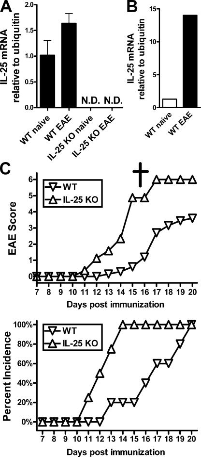
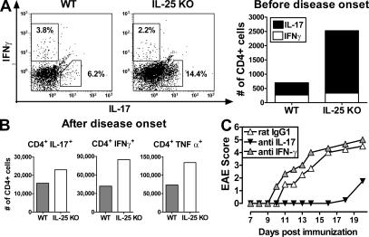
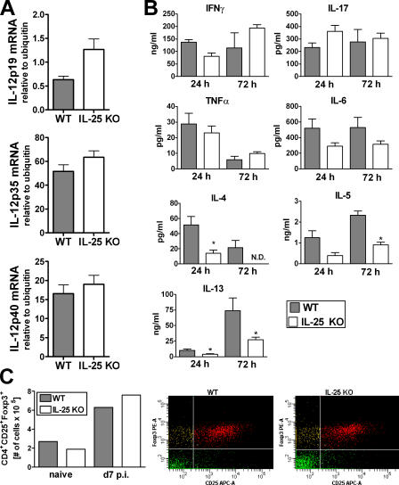
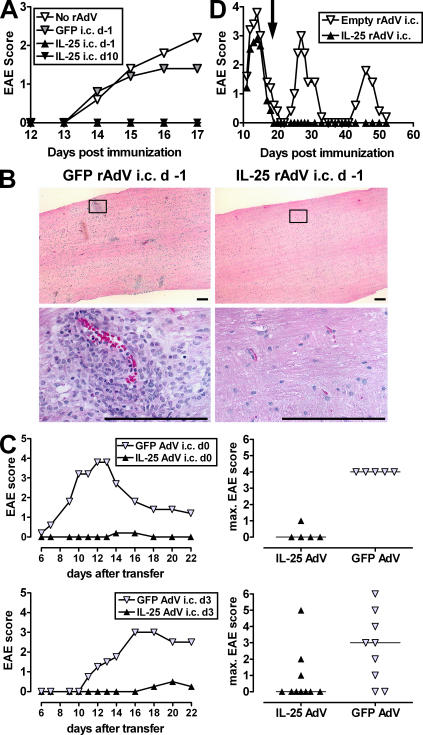
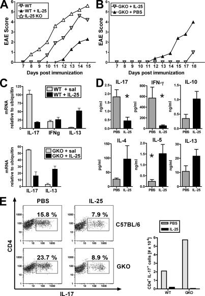
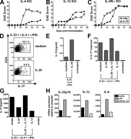
Interleukin (IL)-25 is a member of the IL-17 family of cytokines. However, unlike the other members of this family, IL-25 promotes T helper (Th) 2 responses. We now show that IL-25 also regulates the development of autoimmune inflammation mediated by IL-17-producing T cells. We have generated IL-25-deficient (il25-/-) mice and found that they are highly susceptible to experimental autoimmune encephalomyelitis (EAE). The accelerated disease in the il25-/- mice is associated with an increase of IL-23 in the periphery and a subsequent increase in the number of inflammatory IL-17-, IFNgamma-, and TNF-producing T cells that invade the central nervous system. Neutralization of IL-17 but not IFNgamma in il25-/- mice prevented EAE, suggesting that IL-17 is a major disease-promoting factor. IL-25 treatment at several time points during a relapse-remitting model or chronic model of EAE completely suppressed disease. IL-25 treatment induced elevated production of IL-13, which is required for suppression of Th17 responses by direct inhibition of IL-23, IL-1beta, and IL-6 expression in activated dendritic cells. Thus, IL-25 and IL-17, being members of the same cytokine family, play opposing roles in the pathogenesis of organ-specific autoimmunity.


Figures

Figure 1.
Figure 1.
il25−/− mice are highly susceptible to EAE. (A) Taqman PCR analysis of IL-25 mRNA levels in spinal cords from C57BL/6 WT normal and diseased versus il25-LacZ-KI normal and diseased (n = 6 mice per group). Data shown are representative of at least two experiments. ND, not detectable. (B) IL-25 mRNA expression in CNS-resident microglia. Brain leukocytes were isolated from bone marrow chimeras expressing the CD45.2 allotype on radiation-resistant host cells and the CD45.1 allotype on donor leukocytes. CD11b+CD45.2loCD45.1neg resident microglia were purified from >100 irradiation bone marrow chimeric naive or diseased (day 14) mice. Data from quantitative real-time PCR analysis for IL-25 expression were normalized to ubiquitin. (C) Clinical score (top) and percentage of incidence of disease (bottom) of C57BL/6 (WT) and il25−/− mice immunized with MOG/CFA s.c. on day 0 and PTX i.v. on days 0 and 2. The dagger indicates that by day 17, all il25−/− mice had died from EAE-associated complications. Data shown are 7–9 mice per group and are representative of at least three experiments.
Figure 2.
Figure 2.
Increased numbers of CNS-infiltrating inflammatory Th17 cells in il25−/− mice. (A) Intracellular cytokine staining of FITC-gated CD4+ T cells isolated from the CNS of mice at day 7 after immunization (left, flow cytometric analysis; right, calculated number of cytokine-producing cells). (B) Number of cytokine-producing CD4+ cells in CNS taken at peak of disease. For A and B, cells from four mice were pooled for intracellular cytokine analysis. Data are representative of two experiments. (C) Disease score of il25−/− mice immunized with MOG/CFA (n = 4 mice per group) and treated with antibodies against IL-17 or IFNγ or with rat IgG1 control antibody. Data shown are representative of two experiments.
Figure 3.
Figure 3.
Reduced Th2 cytokine production but normal regulatory T cell response in il25−/− DLN cells. (A) mRNA expression in MOG-stimulated DLN cells taken at day 7 after immunization. Expression levels are normalized to ubiquitin. n = 5. Obtained DLN samples were pooled in three different pools for mRNA processing. (B) In vitro cytokine recall response. DLN cells from MOG-immunized il25−/− and WT mice were isolated at day 7 after immunization. Cells were stimulated with MOG peptide for 3 d and then restimulated with anti-CD3/anti-CD28 for 2 additional days. Supernatants were from 24- or 72-h cultures as indicated. Data are representative of three experiments. All cytokines were measured by cytometric bead array except for IL-13 and IL-17, which were detected by ELISA as described in Materials and methods. ND, not detectable. *, P < 0.05 (unpaired two-tailed t test). (C) Generation of regulatory T cells in il25−/− and WT mice before (pooled sample of two mice per group) and 7 d after (pooled sample of four mice per group) immunization with MOG/CFA. DLN cells were stained for CD4, CD25, and Foxp3.
Figure 4.
Figure 4.
IL-25 inhibits the effector phase of EAE and suppresses clinical disease relapse. (A) Clinical score of SJL mice (n = 5 per group) injected with 3 × 10⁹ particles of indicated rAdV by intracerebral ventricular injection 1 d before or 10 d after immunization. (B) Histopathological analysis of spinal cords from SJL mice at day 18 after immunization. Mice were treated with GFP-rAdV (left) or IL-25-rAdV (right) at day 1. (bottom) 10-fold magnification of boxed area in top panels. Bars, 100 μm. Results of histological analysis are summarized in Table S1 (available at http://www.jem.org/cgi/content/full/jem.20061738/DC1). (C) Clinical score of mice (n = 4–5 per group) that received 2 × 10⁷ activated PLP-specific lymphocytes. Recipient mice were treated with 3 × 10⁹ particles of indicated rAdV at day 0 (top) or day 3 (bottom) after PLP-cell adoptive transfer. (left) Kinetics of mean disease score; (right) maximum disease score per individual mouse during 16 d of observation. (D) Clinical score of EAE-primed mice (n = 7–10 per group) injected with 3 × 10⁹ particles of indicated rAdV by an intracerebral ventricular route during disease remission (20 d after immunization, arrow). Mice were observed for two disease-relapse cycles. Results are representative of at least two experiments.
Figure 5.
Figure 5.
IL-25 induces Th2 and suppresses Th17 independent of IFNγ. (A) Clinical score of immunized WT and il25−/− mice compared with immunized WT mice injected daily s.c. with mIL-25 protein from day 1 to the end of the experiment. Results are representative for at least four experiments with five to six mice per group. (B) Clinical score of ifng−/− (GKO) mice (n = 5 per group) injected daily with PBS or mIL-25 (s.c.) from days 1–12. Results are representative of two experiments. (C) DLN cells from MOG-immunized WT or ifng−/− mice treated with IL-25 or saline were analyzed for cytokine gene expression at day 7 after immunization (five mice per group; DLN cells were processed in three different pools for mRNA analysis). Cytokine gene expression was normalized to ubiquitin. (D) In vitro cytokine recall response. DLN cells from MOG-immunized WT mice treated with PBS or mIL-25 daily s.c. were isolated at day 7 after immunization. Cells were stimulated with MOG peptide for 3 d and restimulated with anti-CD3/anti-CD28 for 2 additional days. Cytokine levels in supernatants were measured by CBA or ELISA. Cells from individual animals were cultured separately, and data are mean cytokine levels ± SD of three to five mice per treatment group. For IFNγ detection, nine mice per treatment group are shown. Data are representative of at least two experiments. *, P < 0.05. (E) DLN cells from MOG-immunized WT and ifng−/− (GKO) mice treated with IL-25 or saline (pool of four mice per group) were stimulated in vitro with MOG for 3 d followed by incubation with IL-2 for another 2 d. Frequencies (FACS plots) and absolute numbers (bar graph) of MOG-specific IL-17–producing CD4+ cells were assessed. Data shown are representative of two experiments.
Figure 6.
Figure 6.
IL-13 is required for Th17 suppression in IL-25–mediated protection from EAE. Clinical score of il4 −/− (A), il13 −/− (B), or il4ra −/− mice (C), with five mice per group injected daily with PBS or mIL-25 (s.c.) from days 1–12. Results are representative of two experiments. (D–F) Naive T cells and LPS-activated CD11c+ DCs were obtained from DO11.10 × RAG−/− mice (pool of five mice) and incubated for 3 d with IL-23 in the presence of OVA peptide. IFNγ and IL-4 were neutralized in these cultures. (D) The effect of IL-25 addition on the number of IL-23–induced IL-17 producers was assessed by intracellular staining for IL-17. (E) IL-13 expression in these cultures was determined by LUMINEX. (F) The modulation of IL-17 protein production in IL-23–driven cultures by IL-25, IL-25 plus anti–IL-13, and IL-13 is shown in percentage of IL-23–induced IL-17 production without addition of exogenous factors (mean IL-17 production is 202.3 pg/ml, assessed by LUMINEX). Data shown are representative of at least two experiments (D and E) or pooled data from two similar experiments (F). (G) Purified MOG33-55 TCR transgenic T cells and LPS-activated CD11c+ DCs were incubated for 4 d with IL-23 in the presence of MOG peptide and neutralizing antibodies against IFNγ and IL-4. IL-17 production after incubation with IL-25, IL-25 plus anti–IL-13, and IL-13 was measured by LUMINEX. (H) DCs were purified from spleens and superficial lymph nodes and activated with LPS with or without addition of IL-13. After 24 h, mRNA expression relative to ubiquitin was measured for IL-23p19, IL-1β, and IL-6 by quantitative real-time PCR. Shown is an experiment using DCs from C57BL/6 mice representative of two C57BL/6 and two BALB/c experiments with a similar outcome (n = 3–5 mice pooled per experiment).

References

    1. Fort, M.M., J. Cheung, D. Yen, J. Li, S.M. Zurawski, S. Lo, S. Menon, T. Clifford, B. Hunte, R. Lesley, et al. 2001. IL-25 induces IL-4, IL-5, and IL-13 and Th2-associated pathologies in vivo. Immunity. 15:985–995. - PubMed
    1. Hurst, S.D., T. Muchamuel, D.M. Gorman, J.M. Gilbert, T. Clifford, S. Kwan, S. Menon, B. Seymour, C. Jackson, T.T. Kung, et al. 2002. New IL-17 family members promote Th1 or Th2 responses in the lung: in vivo function of the novel cytokine IL-25. J. Immunol. 169:443–453. - PubMed
    1. Ikeda, K., H. Nakajima, K. Suzuki, S. Kagami, K. Hirose, A. Suto, Y. Saito, and I. Iwamoto. 2003. Mast cells produce interleukin-25 upon Fc epsilon RI-mediated activation. Blood. 101:3594–3596. - PubMed
    1. Kang, C.M., A.S. Jang, M.H. Ahn, J.A. Shin, J.H. Kim, Y.S. Choi, T.Y. Rhim, and C.S. Park. 2005. Interleukin-25 and interleukin-13 production by alveolar macrophages in response to particles. Am. J. Respir. Cell Mol. Biol. 33:290–296. - PubMed
    1. Owyang, A.M., C. Zaph, E.H. Wilson, K.J. Guild, T. McClanahan, H.R. Miller, D.J. Cua, M. Goldschmidt, C.A. Hunter, R.A. Kastelein, and D. Artis. 2006. Interleukin 25 regulates type 2 cytokine-dependent immunity and limits chronic inflammation in the gastrointestinal tract. J. Exp. Med. 203:843–849. - PMC - PubMed

Publication types

MeSH terms