. 2007 Jan 1;176(1):11-7.
doi: 10.1083/jcb.200605099.

Two binding partners cooperate to activate the molecular motor Kinesin-1

Affiliations

Two binding partners cooperate to activate the molecular motor Kinesin-1

T Lynne Blasius et al. J Cell Biol. .

Abstract

The regulation of molecular motors is an important cellular problem, as motility in the absence of cargo results in futile adenosine triphosphate hydrolysis. When not transporting cargo, the microtubule (MT)-based motor Kinesin-1 is kept inactive as a result of a folded conformation that allows autoinhibition of the N-terminal motor by the C-terminal tail. The simplest model of Kinesin-1 activation posits that cargo binding to nonmotor regions relieves autoinhibition. In this study, we show that binding of the c-Jun N-terminal kinase-interacting protein 1 (JIP1) cargo protein is not sufficient to activate Kinesin-1. Because two regions of the Kinesin-1 tail are required for autoinhibition, we searched for a second molecule that contributes to activation of the motor. We identified fasciculation and elongation protein zeta1 (FEZ1) as a binding partner of kinesin heavy chain. We show that binding of JIP1 and FEZ1 to Kinesin-1 is sufficient to activate the motor for MT binding and motility. These results provide the first demonstration of the activation of a MT-based motor by cellular binding partners.


Figures
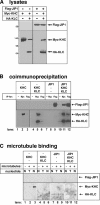
Figure 1.
Figure 1.
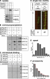
Binding of JIP1 to Kinesin-1 is not sufficient for activation. (A) COS cell lysates expressing the indicated proteins were immunoblotted with antibodies to the Flag (top) or myc and HA tags (bottom). (B) The indicated lysates were immunoprecipitated (IP) with antibodies to the myc or Flag tags or no antibody as a control (−). Precipitates were immunoblotted with antibodies to the Flag tag (top) or myc and HA tags (bottom). (C) Prepolymerized taxol-stabilized MTs were added (+) or not added (−) to the indicated lysates with either ATP (T) or AMPPNP (N). After sedimentation, the MT pellets were immunoblotted with antibodies to the Flag (top) or myc and HA tags (bottom). Panels are from different parts of the same gel.
Figure 2.
Figure 2.
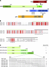
FEZ1 binds to the inhibitory tail of KHC. (A) Schematic illustration of the domain structures of FEZ1, KHC, KLC, and JIP1. Interacting regions are indicated with a double arrow. TPR, tetritricopeptide repeat; PTB, phosphotyrosine binding. (B) Sequence alignment of KHC tail domains from Homo sapiens (Hs; GenBank/EMBL/DDBJ accession no. BAA25457), Drosophila (Dm; P17210), Caenorhabditis elegans (Ce; AAA28155), and N. crassa (Nc; P48467). Sequences were aligned using T-Coffee (Notredame et al., 2000), converted to Clustal-W, and modified by ESPript 2.2. Green overline, conserved coiled tail implicated in cargo binding; black overline, globular tail implicated in autoinhibition; dashed underline, folding site; asterisks, IAK sites; white text highlighted in red, identical residues; red text on white background, conserved residues. (C) Ability of truncated or mutant versions of KHC(750–955) to interact with FEZ1.
Figure 3.
Figure 3.
FEZ1 and JIP1 cooperate to activate Kinesin-1 in vitro. (A) COS cell lysates expressing the indicated proteins were immunoblotted with antibodies to the Flag tag (top) or to FEZ1 and the myc and HA tags (bottom). (B) The indicated lysates were mixed and immunoprecipitated (IP) with antibodies to KHC or the Flag tag or to no primary antibody as a control (−). Precipitates were immunoblotted with antibodies to the Flag tag (top), FEZ1 (middle), or myc and HA tags (bottom). (C) MT-binding assay as in Fig. 1 C. MT pellets were immunoblotted with antibodies to the Flag tag (top), FEZ1 (middle), or myc and HA tags (bottom). T, ATP; N, AMPPNP. (B and C) Panels are from different parts of the same gel. (D–F) Single-molecule motility assay. (D) Myc-KHC + 3xmCit-KLC lysates were mixed with lysates of mock-transfected cells (left) or cells expressing Flag-JIP1 and FEZ1-hsv (right). Representative motile events along Cy5-labeled MTs are shown in the kymographs (13 frames; 100-ms intervals). Bar, 1.0 μm. (E) Speed histogram. Myc-KHC + 3xmCit-KLC motors activated by JIP1 and FEZ1 move at a mean speed of 0.71 ± 0.31 μm/s (n = 47). Data are pooled from three separate experiments. (F) Single exponential decay fitting (red line) of the run length histogram shows that the same motors move processively for 0.46 ± 0.05 μm/run.
Figure 4.
Figure 4.
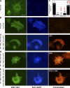
FEZ1 and JIP1 cooperate to activate Kinesin-1 in live cells. (A–E) COS cells expressing the indicated proteins were exposed to AMPPNP for 10 min and were fixed and stained. Bar, 25 μm. (F) Fluorescence relocation index of Kinesin-1 over time was determined from videos (available at http://www.jcb.org/cgi/content/full/jcb.200605099/DC1) taken during permeabilization and AMPPNP treatment. n values in parentheses indicate the number of videos. Error bars represent SD. #, P > 0.5; **, P < 0.001 compared with KHC + KLC.
Figure 5.
Figure 5.
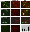
Dominant-negative FEZ1 disrupts Kinesin-1 transport of JIP1. (A–D) Differentiated CAD cells expressing myc-FEZ1 (A), myc-FEZ1(1–230; B), or myc-FEZ1(1–308; C) or untransfected cells (D) were double labeled for the myc tag and endogenous JIP1 protein. Arrows indicate tips of neurites in transfected cells. Bar, 10 μm. (E) Quantification of JIP1 tip staining. n values in parentheses indicate the number of cells. Error bars represent SD. *, P ≈ 0.01; **, P < 0.001 compared with untransfected cells.

Comment in

  • Jump-starting kinesin.
    Hackney DD. Hackney DD. J Cell Biol. 2007 Jan 1;176(1):7-9. doi: 10.1083/jcb.200611082. J Cell Biol. 2007. PMID: 17200413 Free PMC article.

Similar articles

Cited by

References

    1. Adio, S., J. Reth, F. Bathe, and G. Woehlke. 2006. Review: regulation mechanisms of Kinesin-1. J. Muscle Res. Cell Motil. 27:153–160. - PubMed
    1. Bathe, F., K. Hahlen, R. Dombi, L. Driller, M. Schliwa, and G. Woehlke. 2005. The complex interplay between the neck and hinge domains in kinesin-1 dimerization and motor activity. Mol. Biol. Cell. 16:3529–3537. - PMC - PubMed
    1. Bloom, L., and H.R. Horvitz. 1997. The Caenorhabditis elegans gene unc-76 and its human homologs define a new gene family involved in axonal outgrowth and fasciculation. Proc. Natl. Acad. Sci. USA. 94:3414–3419. - PMC - PubMed
    1. Cai, D., A.D. Hoppe, J.A. Swanson, and K.J. Verhey. 2007. Kinesin-1 structural organization and conformational changes revealed by FRET stoichiometry in live cells. J. Cell Biol. 176:51–63. - PMC - PubMed
    1. Coy, D.L., W.O. Hancock, M. Wagenbach, and J. Howard. 1999. Kinesin's tail domain is an inhibitory regulator of the motor domain. Nat. Cell Biol. 1:288–292. - PubMed

Publication types

MeSH terms