. 2007 Jan 1;176(1):27-33.
doi: 10.1083/jcb.200609116.

Two distinct modes of myosin assembly and dynamics during epithelial wound closure

Affiliations

Two distinct modes of myosin assembly and dynamics during epithelial wound closure

Masako Tamada et al. J Cell Biol. .

Erratum in

  • J Cell Biol. 2007 Feb 12;176(4):545

Abstract

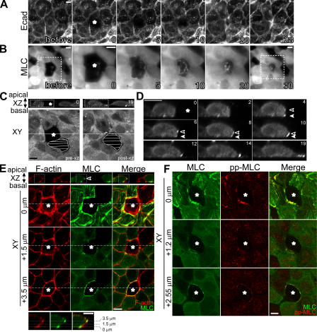
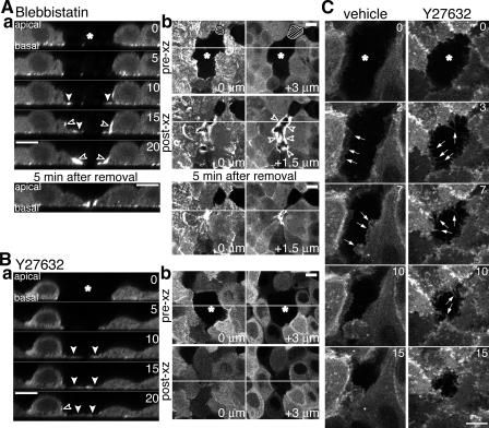
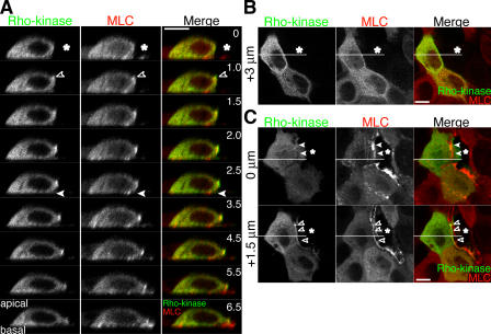
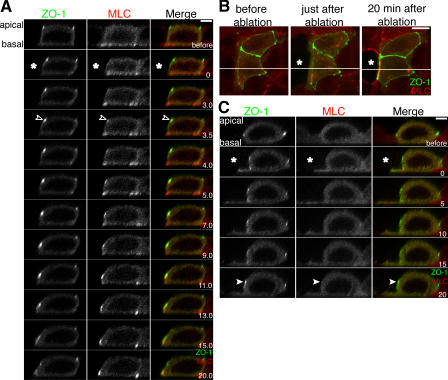
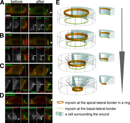
Actomyosin contraction powers the sealing of epithelial sheets during embryogenesis and wound closure; however, the mechanisms are poorly understood. After laser ablation wounding of Madin-Darby canine kidney cell monolayers, we observed distinct steps in wound closure from time-lapse images of myosin distribution during resealing. Immediately upon wounding, actin and myosin II regulatory light chain accumulated at two locations: (1) in a ring adjacent to the tight junction that circumscribed the wound and (2) in fibers at the base of the cell in membranes extending over the wound site. Rho-kinase activity was required for assembly of the myosin ring, and myosin II activity was required for contraction but not for basal membrane extension. As it contracted, the myosin ring moved toward the basal membrane with ZO-1 and Rho-kinase. Thus, we suggest that tight junctions serve as attachment points for the actomyosin ring during wound closure and that Rho-kinase is required for localization and activation of the contractile ring.


Figures

Figure 1.
Figure 1.
The dynamics of MLC localization and cell shape changes during wound closure. (A and B) One or two cells in a monolayer of MDCK cells expressing Ecad-RFP (A) or MLC-EGFP (B) were laser ablated. Epifluorescent images of cells were collected before and after laser ablation. Asterisks indicate the ablated cells. Elapsed time is indicated on each panel in minutes. Micrographs of 0, 5, 10, and 20 min in B show the images of the boxed region at higher magnification. (C) One cell in MLC-EGFP cell monolayer was laser ablated, and time-lapse XZ images of MLC-EGFP were collected for 19 min. XY sections were taken when XZ imaging was started (pre-xz) and finished (post-xz). White lines in XY sections indicate the position of XZ sections. Hatched regions represent cells with low MLC-EGFP. (D) Time-lapse images of the boxed region in C at higher magnification. MLC-EGFP accumulated at the apical–lateral border (open arrowheads) and basal–lateral border (closed arrowheads) of cells surrounding the ablated cell. (E) About 5 min after ablation, MLC-EGFP cells were fixed and F-actin was visualized with Alexa Fluor 568 phalloidin. The bottom panels show the images of the boxed regions at higher magnification. MLC-EGFP concentrated into a ring at the apical–lateral border and into fibers at the basal–lateral border with F-actin around wound. (F) Phospho-MLC accumulates around the wound. MLC-EGFP cells were treated and fixed as in E and stained with anti–phospho-MLC (Thr18/Ser19) antibody. Protein distributions are shown at three Z positions. Bars, 10 μm.
Figure 2.
Figure 2.
Myosin activity is required for contraction of the actomyosin ring and wound closure, and Rho-kinase is involved in ring accumulation. MLC-EGFP cells were treated with 50 μM blebbistatin for 20 min (A) or 20 μM Y27632 for 1 h (B) before laser ablation. After ablation, cells were observed for 20 min in the presence of inhibitors (a). (A) In the presence of blebbistatin, MLC-EGFP accumulated, but the wound remained open 20 min after laser ablation (open arrowheads). Basal membranes extended into the wound space (closed arrowheads). 5 min after removal of blebbistatin, the wound was closed (a and b, bottom). (B) In the presence of Y27632, the accumulation of MLC-EGFP at the apical–lateral border was attenuated (open arrowhead), but basal membranes extended into the wound space (closed arrowheads). (C) After ablation, the basal plane of EGFP-actin cells was observed. Cells extended into the wound site with lamellipodial protrusions (left, arrows), and these lamellipodial protrusions were replaced by filopodia in the presence of Y27632 (right, arrows). Asterisks indicate the ablated cells. Bars, 10 μm.
Figure 3.
Figure 3.
Rho-kinase accumulates at the apical–lateral border of cells surrounding the wound. (A) Cells in a monolayer of MLC-RFP cells transiently expressing EGFP–Rho-kinase were ablated, and XZ images were collected. (B and C) XY images were taken before (B) and after (C) XZ imaging. EGFP–Rho-kinase and MLC-RFP coaccumulated simultaneously at the apical–lateral border (open arrowheads). EGFP–Rho-kinase did not accumulate at the basal–lateral border (closed arrowheads). Asterisks indicate the ablated cells. Bars, 10 μm.
Figure 4.
Figure 4.
The myosin ring forms at the tight junction. (A) Cells in a monolayer of MLC-RFP cells transiently expressing ZO-1–EGFP were ablated, and XZ images were collected. MLC-RFP started to accumulate at site containing ZO-1–EGFP concentration (open arrowheads), and the two moved together thereafter. (B) Z stacks of XY sections taken before and after ablation were projected along the Z axis, and images of MLC-RFP (red) and ZO-1-EGFP (green) were merged. (C) Cells were treated and observed as in A in the presence of Y27632. MLC-RFP accumulation at the apical–lateral border was attenuated, and ZO-1–EGFP concentration remained at the initial position (closed arrowheads). Asterisks indicate the ablated cells. Bars: (B) 10 μm; (A and C) 5 μm.
Figure 5.
Figure 5.
Comparison between distribution of junctional proteins and distribution of MLC after ablation. (A–D) XZ sections of MLC-EGFP cells expressing Ecad-RFP (A) and MLC-RFP cells expressing EGFP–α-catenin (B), EGFP–l-afadin (C), or EGFP-claudin1 (D) were observed before (left) and after (right) ablation. Bottom panels show the boxed regions of the top panel at higher magnification. Arrows indicate the sites of MLC accumulation at the apical–lateral border. Asterisks indicate the ablated cells. Bars, 5 μm. (E) Diagram of myosin accumulation and its dynamics during wound closure.

Similar articles

Cited by

References

    1. Bement, W.M., P. Forscher, and M.S. Mooseker. 1993. A novel cytoskeletal structure involved in purse string wound closure and cell polarity maintenance. J. Cell Biol. 121:565–578. - PMC - PubMed
    1. Benink, H.A., and W.M. Bement. 2005. Concentric zones of active RhoA and Cdc42 around single cell wounds. J. Cell Biol. 168:429–439. - PMC - PubMed
    1. Bertet, C., L. Sulak, and T. Lecuit. 2004. Myosin-dependent junction remodelling controls planar cell intercalation and axis elongation. Nature. 429:667–671. - PubMed
    1. Ehrlich, J.S., M.D. Hansen, and W.J. Nelson. 2002. Spatio-temporal regulation of Rac1 localization and lamellipodia dynamics during epithelial cell-cell adhesion. Dev. Cell. 3:259–270. - PMC - PubMed
    1. Ikebe, M., and D.J. Hartshorne. 1985. Phosphorylation of smooth muscle myosin at two distinct sites by myosin light chain kinase. J. Biol. Chem. 260:10027–10031. - PubMed

Publication types

Substances