. 2006 Dec 9:12:1565-85.

Interphotoreceptor retinoid-binding protein gene structure in tetrapods and teleost fish

Affiliations

Interphotoreceptor retinoid-binding protein gene structure in tetrapods and teleost fish

John M Nickerson et al. Mol Vis. .

Abstract

Purpose: The interphotoreceptor retinoid-binding protein (IRBP) gene possesses an unusual structure, encoding multiple Repeats, each consisting of about 300 amino acids. Our goals were to gain insight into the function of IRBP, and to test the current model for the evolution of IRBP, in which Repeats were replicated from a simpler ancestral gene.

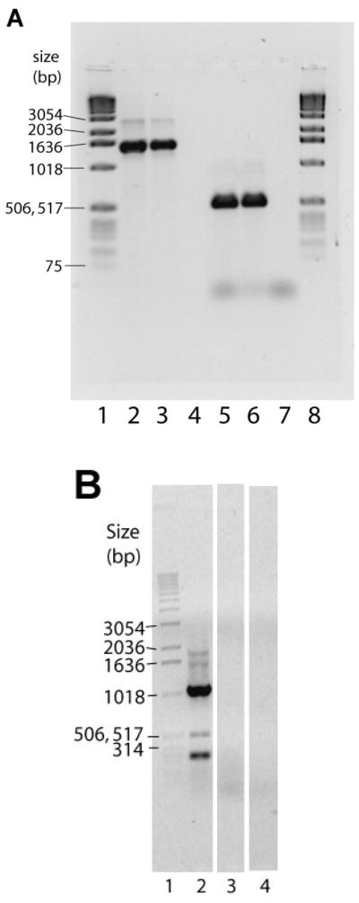
Methods: We employed a bioinformatics approach to analyze IRBP loci in recently completed or near-complete genome sequences of several vertebrates and nonvertebrate chordates. IRBP gene expression in zebrafish was evaluated by reverse transcriptase PCR (RT-PCR) and in situ mRNA hybridizations with gene-specific probes.

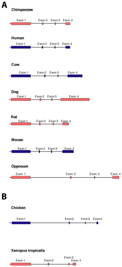
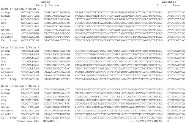
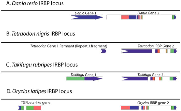
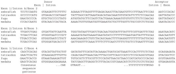
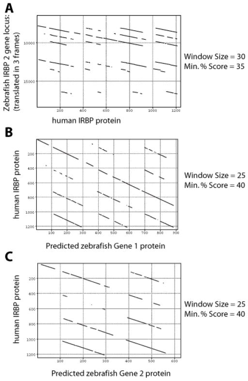
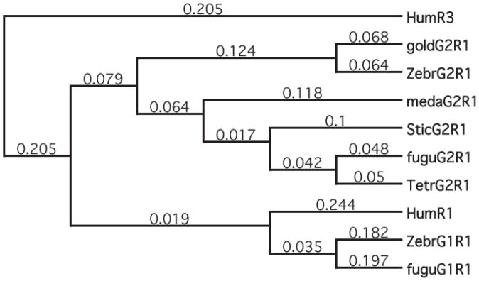
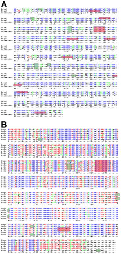
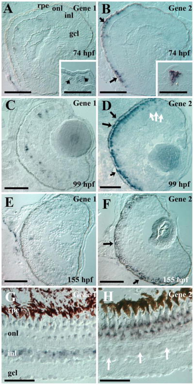
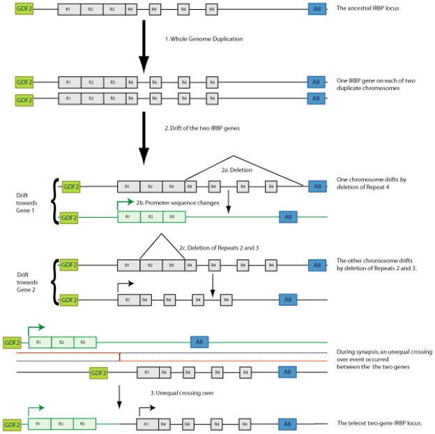
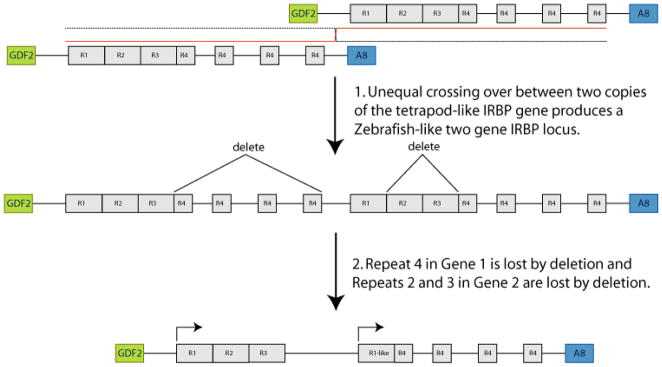
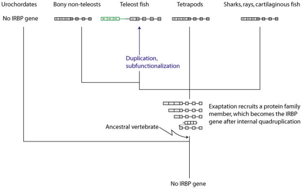
Results: Patterns of exons and introns in the IRBP genes of tetrapods were highly similar, as were predicted amino acid sequences and Repeat structures. IRBP gene structure in teleost fish was more variable, and we report a new gene structure for two species, the Japanese puffer fish (Takifugu rubripes) and the zebrafish (Danio rerio). These teleost genomes contain a two-gene IRBP locus arranged head-to-tail in which the first gene, Gene 1, is intronless and contains a single large exon encoding three complete Repeats. It is followed by a second gene, Gene 2, which corresponds to the previously reported gene consisting of two Repeats spread across four exons and three introns. Each of the two zebrafish genes is transcribed. Gene 2 is expressed in the photoreceptors and RPE, and Gene 1 is expressed in the inner nuclear layer and weakly in the ganglion cell layer.

Conclusions: The tetrapod IRBP gene structure is highly conserved while the teleost fish gene structure was a surprise: It appears to be a two-gene locus with distinct Repeat organization in each open reading frame. This gene structure and gene expression data are consistent with possible neofunctionalization or sub-function partitioning of Gene 1 and Gene 2 in the zebrafish. We suggest that the two-gene locus in teleost fish arose as a consequence of either the known whole genome duplication or single gene tandem duplication.


Figures

Figure 1
Figure 1
Tetrapod IRBP gene structures. There is a common gene structure for IRBP among the mammals, birds, and amphibians with a large first exon encoding three full Repeats and parts of the fourth and final Repeat are spread among the first through fourth exons. The blue genes are previously published (human [66,67], mouse [48], cow [26], and chicken [51]), while the red genes are mined from data made available thorough the courtesy of several large-scale sequencing centers as identified in the Acknowledgments. A illustrates the gene structures of the mammals. The dog IRBP gene may have an extra Intron very near the 3′ end of the gene, and this is in the 3′ UTR. Among the orthologs, the positions of the introns in IRBP are constant, and the coding regions in each exon are the same. B shows the IRBP gene structures from non-mammalian tetrapods.
Figure 2
Figure 2
Alignment and comparison of Donor and Acceptor sites from the tetrapods. This is a comparison to the accepted consensus donor and acceptor sites from human genes. All the introns contain invariant GT and AG dinucleotides at the beginning and end of the intron. Most nearby nucleotides closely match the consensus donor or acceptor motif. The lariat consensus sequence is detected in about half the introns within 50 nucleotides of the acceptor site, and all have several A’s, which may function in the absence of a closely matching lariat sequence. These alignments indicate that the splice sites are all strongly conserved in position and sequence, and they all appear to function well.
Figure 3
Figure 3
Teleost fish IRBP gene loci. A: Zebrafish has two IRBP genes. B: Tetraodon nigroviridis has a remnant of the upstream gene (Gene 1) and a full copy of Gene 2. C: Fugu has two IRBP genes. Panel D: Medaka has a single gene corresponding to Gene 2. In zebrafish and fugu, the two IRBP genes are oriented head to tail, and, across species, the intergenic spacer is about the same size. No intervening genes are found between the two genes in the single locus. The colors originate with the web site computer program called AUGUSTUS [46]. Each exon is marked with two different colors, such that the end of one exon and the beginning of the next exon have the same color. Colors are rotated so that adjacent exons are colored differently (only three colors are needed) and the order of use is blue-red-green, which is then repeated. The first exon of a gene is depicted with an arrow tail at the 5′ end, and the last exon is labeled with an arrowhead at the 3′ end. For genes that have only a single exon, they are solid blue, with an arrow head and tail. These models are predicted gene structures based on several algorithms. Also, some of the features are small, so that a tiny first exon might not appear to have an arrow tail at the 5′ end.
Figure 4
Figure 4
Alignment and comparison of Donor and Acceptor sites from the teleost fish Gene 2. This is a comparison to the accepted consensus donor and acceptor sites from human genes. At the beginning and end of each intron, respectively, invariant GT and AG dinucleotides are located. Most nearby nucleotides match the consensus donor or acceptor motif. These alignments indicate that the splice sites are all strongly conserved in position and sequence, and they all appear to function correctly.
Figure 5
Figure 5
Dot matrix comparisons showing the existence of a two-gene IRBP locus in zebrafish. A: Dot matrix of human protein against the entire zebrafish gene locus. B: Human protein against translated Gene 1. C: Human protein against Gene 2 protein. The Pustell dot matrix program in MacVector was used employing the pam250 weight matrix and a hash value of 2 in all three panels. Two genes exist in tandem. The genes are oriented head to tail, the genes are close together, the genes are different, with Gene 1 a single exon and three-Repeats long and Gene 2 having four exons, but just two repeats. The optimum alignments to the human IRBP amino acid sequence showed that Gene 1 encodes Repeats 1, 2, and 3, while Gene 2 encoded Repeats 1 and 4.
Figure 6
Figure 6
Phylogenetic analysis of Repeat 1 from IRBP. Evolutionary relationships among fish and human Repeat 1 orthologs and paralogs are illustrated. MacVector version 9.0 was used to build a cladogram of the Repeat 1-like amino acid sequences of the teleost fish and the human Repeat 1. Human Repeat 3 was used as an outgroup for the purpose of rooting this tree. The illustrated tree was built using the neighbor-joining method. Whether using the UPGMA or neighbor-joining method, with uncorrected or Poisson-corrected distances, random or systematic tie-breaking for gaps, in MacVector version 9.0, all the phylogenetic reconstructions gave the same overall structure. A consensus tree from 1000 replications of bootstrapping was calculated, and it was identical to the structure of the best tree, which is illustrated in this image. Among 1000 replications of resampling, all seven relevant nodes occurred frequently, with four appearing in 100% of trees, two nodes occurring in 96% of trees, and one node occurring in 79% of trees. All the nodes are resolved in the illustrated tree. The results suggest a paralogous relationship between the Repeat 1 sequences from Genes 1 and 2 in zebrafish and fugu. The closer relationship of human Repeat 1 to the fugu and zebrafish Gene 1 orthologs than to Gene 2 and the illustrated divergence at the leftmost internal node of the paralogs on this cladogram suggests that the paralogous Genes 1 and 2 arose early or at the origin of the teleosts. This tree is consistent with the already known divergence radiation of the teleost fish about 350 Mya.
Figure 7
Figure 7
Multiple alignment of fish amino acid sequences. A: Gene 1 from zebrafish (zebr1) and fugu (fugu1) are aligned over Repeats 1 through 3, about 930 amino acids. B: Gene 2 Repeats 1 and 2 orthologs from fugu, goldfish, medaka, stickleback, tetraodon, and zebrafish are aligned over about 620 amino acids. Motifs identified: Signal peptide (leader sequence) cleavage sites are shown by the arrow. Glycosylation sites, NX(T|S), are shown in gray boxes with a transparent green fill. Hyaluronan binding sites, (R|K)X7(R|K), are shown in a blue box with a transparent light red fill. Identical amino acids across all aligned positions are shown in blue. Red illustrates identical residues in 3 to 5 of the 6 possible identities. Green illustrates residues with similar chemical properties at a given position.
Figure 8
Figure 8
RT-PCR from Gene 1 or Gene 2-specific primers. A: Expression of Gene 1 and Gene 2 in zebrafish tissues. Amplicon size was measured on a 1.0% agarose gel (and run in 1X TAE buffer) stained with 1X SYBR Green. Lanes 1 and 8, 0.5 μg 1 kb DNA ladder (Invitrogen, Carlsbad, CA). Lane 2, 96 h zebrafish whole larval RNA primed with gn1fwd and gn1rev (Table 2). Lane 3, adult zebrafish eye RNA primed with gn1fwd and gn1rev primers. Lane 4, no RNA control, primed with gn1fwd and gn1rev. Lane 5, 96 h whole larval RNA with gn2 primers, F3 and R4 (Table 2). Lane 6, adult zebrafish eye RNA, primed with gn2 primers, F3 and R4. In the absence of RNA, the F3 and R4 primers amplified no products (data not shown). Lanes 2 and 3 had a product of about 1500 bp, and the size expected from the Gene 1 DNA sequence was 1529 bp. Lanes 5 and 6 had a product of about 530 bp and an expected size based on the sequence of 532 bp. These results indicate that both genes are transcriptionally active in both zebrafish adult eye and larval whole bodies. Panel B: Expression of an RNA transcript spanning Gene 1 and Gene 2. Lane 1, 1 kb ladder (Invitrogen). Lane 2, an RT-PCR product spanning across the intergenic region using primers F1 and R3 (Table 1) was found with a size estimated to be 1000 bp (96 h larval RNA). Lane 3, same as Lane 2 in the absence of reverse transcriptase. Lane 4, no RNA control.
Figure 9
Figure 9
In situ hybridizations. Zebrafish IRBP Gene 1 and Gene 2 are differentially expressed. Retinal cryosections were obtained from 74 hpf (A, B) albb4 zebrafish embryos, 99 hpf (C, D) and 155 hpf (E, F) albb4 zebrafish larvae, and adult Tue zebrafish eyes (G, H) and were hybridized with Gene 1- (A, C, E, G) and Gene 2- (B, D, F,H) specific probes. Note that albb4 zebrafish do have low levels of pigment in the RPE; dark arrows show Gene 2 hybridization. White arrows show weak expression of Gene 2 sporadically in the INL. Insets in A and B shows Gene 1 and Gene 2 expression in developing pineal; contrast was enhanced to show weak expression of Gene 1. Scale bars represent 50 μm; rpe represent retinal pigmented epithelium, onl represent outer nuclear layer; inl represent inner nuclear layer; gcl represent ganglion cell layer.
Figure 10
Figure 10
A model of the origin of two IRBP genes in the teleosts. The presumptive ancestral gene in fish, predating the appearance of the teleosts is a gene much like the present day tetrapod gene with four Repeats encoded in a four-exon gene, with the first three repeats and part of the fourth found in the first exon. When the teleosts arose in Step 1, the whole genome was duplicated. In Step 2, the two IRBP genes drifted, deleting different parts of the gene. In the gene destined to become Gene 1, the 3′ end of the gene was lost deleting the three introns, exons 2–4 and part of exon 1 (Step 2a), and the promoter underwent some alterations (Step 2b), so that it would be expressed in some ganglion cells and selected cells in the inner nuclear layer. The gene destined to become Gene 2 underwent an internal deletion, losing the parts of Exon 1 that encoded Repeats 2 and 3 (Step 2c) but Gene 2 did not undergo any significant changes in its promoter, retaining expression in photoreceptor cells and RPE cells. In Step 3, unequal crossing over between the two chromosomes would form the current teleost two-gene locus. Synteny is preserved in this model. GDF2 represents the growth differentiation factor 2 gene, and A8 represents the Annexin 8 gene.
Figure 11
Figure 11
A model of teleost IRBP locus evolution based on external unequal crossing over. An unequal crossing over event between the two chromatids of the IRBP gene leads to one chromatid bearing two copies of the IRBP gene. In subsequent steps, Repeat 4 is lost from the first gene and Repeats 2 and 3 are lost from the second gene. These steps produce a two-gene locus in head-to-tail orientation. Synteny is preserved in this model. GDF2 represents the growth differentiation factor 2 gene, and A8 represents the Annexin 8 gene.
Figure 12
Figure 12
Current model of the origins and evolution of the IRBP gene. The IRBP gene probably arose coincident with or shortly after the vertebrates diverged from the urochordates. This is the time period when the neural crest and the skull originated. No major changes are proposed in the steps resulting in the early internal quadruplication of the IRBP gene, but the time frame is now bounded, as the IRBP gene appears to be absent from the urochoradates. This model is based on Borst et al. [26] and Rajendran et al. [27]. The major change is the addition of a two-gene IRBP locus in the teleosts.

References

    1. Bunt-Milam AH, Saari JC. Immunocytochemical localization of two retinoid-binding proteins in vertebrate retina. J Cell Biol. 1983;97:703–12. - PMC - PubMed
    1. Crouch RK, Hazard ES, Lind T, Wiggert B, Chader G, Corson DW. Interphotoreceptor retinoid-binding protein and alpha-tocopherol preserve the isomeric and oxidation state of retinol. Photochem Photobiol. 1992;56:251–5. - PubMed
    1. Gonzalez-Fernandez F. Evolution of the visual cycle: the role of retinoid-binding proteins. J Endocrinol. 2002;175:75–88. - PubMed
    1. Liou GI, Wang M, Matragoon S. Timing of interphotoreceptor retinoid-binding protein (IRBP) gene expression and hypomethylation in developing mouse retina. Dev Biol. 1994;161:345–56. - PubMed
    1. Gonzalez-Fernandez F, Van Niel E, Edmonds C, Beaver H, Nickerson JM, Garcia-Fernandez JM, Campohiaro PA, Foster RG. Differential expression of interphotoreceptor retinoid-binding protein, opsin, cellular retinaldehyde-binding protein, and basic fibroblastic growth factor. Exp Eye Res. 1993;56:411–27. Erratum in: Exp Eye Res 1993 Jul;57(1):127. - PubMed

Publication types

MeSH terms

Substances

LinkOut - more resources