Review
. 2007 Jan;117(1):6-12.
doi: 10.1172/JCI30974.

Tales from the crypts: regulatory peptides and cytokines in gastrointestinal homeostasis and disease

Affiliations
Review

Tales from the crypts: regulatory peptides and cytokines in gastrointestinal homeostasis and disease

Juanita L Merchant. J Clin Invest. 2007 Jan.

Abstract

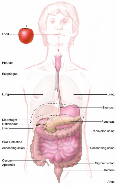
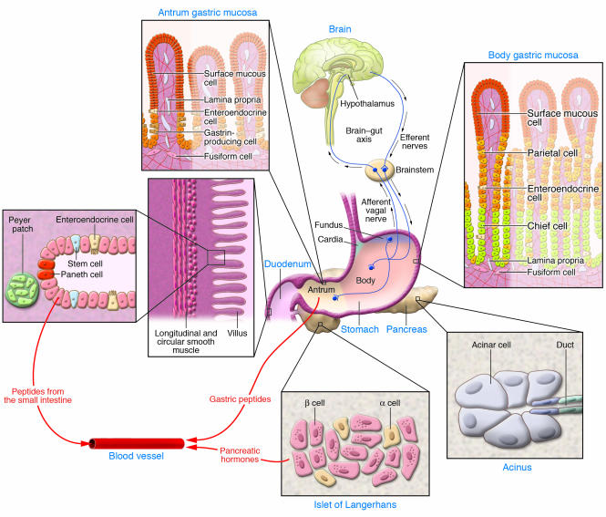
The gastrointestinal (GI) tract is composed of a diverse set of organs that together receive extracorporeal nutrition and convert it to energy substrates and cellular building blocks. In the process, it must sort through all that we ingest and discriminate what is useable from what is not, and having done that, it discards what is "junk." To accomplish these many and varied tasks, the GI tract relies on endogenous enteric hormones produced by enteroendocrine cells and the enteric nervous system. In many instances, the mediators of these tasks are small peptides that home to the CNS and accessory gut organs to coordinate oral intake with digestive secretions. As the contents of ingested material can contain harmful agents, the gut is armed with an extensive immune system. A breach of the epithelial barrier of the GI tract can result in local and eventually systemic disease if the gut does not mount an aggressive immune response.


Figures

Figure 1
Figure 1. Overview of the GI tract.
In humans, the GI tract is a 20-foot muscular tube lined with primarily endodermally derived columnar epithelial cells. The esophagus begins at the end of the posterior pharynx. The esophagus courses through the thoracic cavity and pierces the diaphragm, where it enters the abdominal cavity and merges with the stomach. The stomach merges distally with the small intestine, which is divided into 3 parts, the duodenum, jejunum, and ileum. The terminal ileum joins the colon at the ileocecal valve. The colon begins in the right lower abdomen (ascending colon) and transverses under the liver into the transverse, then left, colon. Just before entering the rectum, the distal left colon forms an S shape (the sigmoid colon). The luminal GI tract ends at the anus. The liver, pancreas, and gallbladder are accessory organs that form as appendages from the second portion of the duodenum.
Figure 2
Figure 2. Components of the neural, endocrine, and immune systems that regulate GI function.
Essential components discussed in this overview are shown. Vagal afferents emerge from the ganglia between the layers of smooth muscle (circular and longitudinal) surrounding the stomach and small intestine to synapse in the spinal cord and brain (collectively the CNS). In addition, the brain (especially the hypothalamus) relays efferent signals to target organs. The human stomach is divided into 4 regions (cardia, fundus, body [corpus], and antrum). The acid-secreting parietal cells are found in the corpus, whereas the gastrin-producing cells are found in the antrum. Neuroendocrine cells secrete gastric peptides such as ghrelin in the corpus and gastrin in the antrum into the circulation through local capillaries. Similarly, regulatory peptides from neuroendocrine cells in the small intestine and pancreatic islets of Langerhans are secreted into the bloodstream. Collections of immune cells are most prominent in the small intestine as Peyer patches. In addition, the Paneth cells at the base of the intestinal crypts secrete antimicrobial peptides including lysozyme and defensins. Stem cells in the small intestine are anatomically well defined as the 4th cell from the base of the crypt.

Similar articles

Cited by

References

    1. Gilbert, S.F. 1994.Developmental biology. Sinauer Associates Inc. Sunderland, Massachusetts, USA. 361–368.
    1. Ahlman H., Nilsson The gut as the largest endocrine organ in the body. Ann. Oncol. 2001;12(Suppl. 2):S63–S68. - PubMed
    1. Kelly D., Coutts A.G. Early nutrition and the development of immune function in the neonate. Proc. Nutr. Soc. 2000;59:177–185. - PubMed
    1. Goyal R.K., Hirano I. The enteric nervous system. N. Engl. J. Med. 1996;334:1106–1115. - PubMed
    1. Wilson, K.H. 1999. The gastrointestinal biota. InTextbook of gastroenterology. T. Yamada, D.H. Alpers, L. Laine, C. Owyang, and D. Powell, editors. Lippincott Williams & Wilkins. Philadelphia, Pennsylvania, USA. 624–636.

Publication types