Review
. 2007 Jan;117(1):50-9.
doi: 10.1172/JCI30082.

The pancreatic stellate cell: a star on the rise in pancreatic diseases

Affiliations
Review

The pancreatic stellate cell: a star on the rise in pancreatic diseases

M Bishr Omary et al. J Clin Invest. 2007 Jan.

Abstract

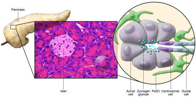
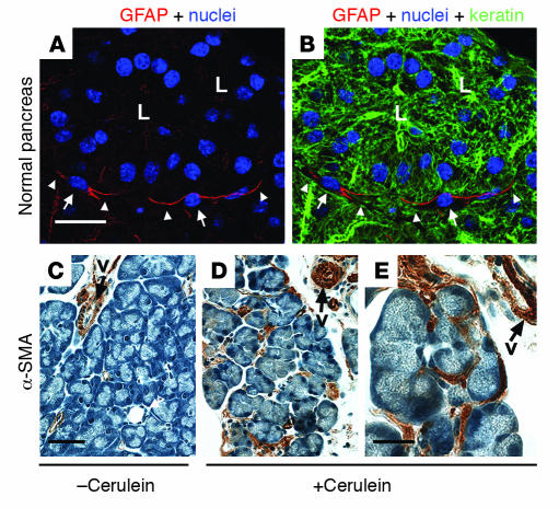
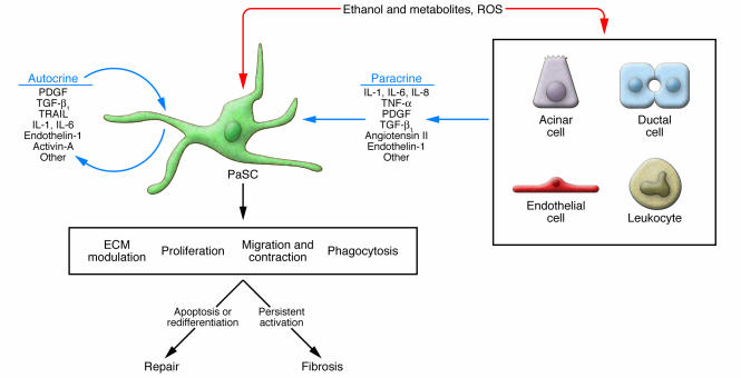
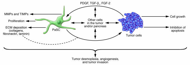
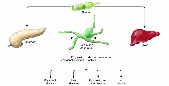
Pancreatic stellate cells (PaSCs) are myofibroblast-like cells found in the areas of the pancreas that have exocrine function. PaSCs are regulated by autocrine and paracrine stimuli and share many features with their hepatic counterparts, studies of which have helped further our understanding of PaSC biology. Activation of PaSCs induces them to proliferate, to migrate to sites of tissue damage, to contract and possibly phagocytose, and to synthesize ECM components to promote tissue repair. Sustained activation of PaSCs has an increasingly appreciated role in the fibrosis that is associated with chronic pancreatitis and with pancreatic cancer. Therefore, understanding the biology of PaSCs offers potential therapeutic targets for the treatment and prevention of these diseases.


Figures

Figure 1
Figure 1. Schematic of the cellular components of the exocrine pancreas.
The pancreas can be functionally divided into 2 components that are interspersed: an exocrine component that consists primarily of acini — clusters of acinar cells that feed into ductules — and an endocrine component composed of the islets. In the normal pancreas, quiescent PaSCs are present in the periacinar space. These cells have long cytoplasmic processes that encircle the base of the acinus. Zymogen granules release their contents of digestive enzymes into the pancreatic ductal system upon stimulation.
Figure 2
Figure 2. Immune staining of PaSCs.
(A and B) Normal mouse pancreas was triple stained to visualize GFAP (red), nuclei (blue), and keratin polypeptide 8 (green). L, lumen; arrows point to PaSC nuclei and arrowheads point to PaSC processes. (CE) Pancreata from plasminogen-deficient mice injected with saline (C) or with cerulein to induce pancreatitis (D and E) were stained with antibodies specific for α-SMA as described previously (61). The image shown in E is a higher magnification of that shown in D. Note the dramatic induction of α-SMA in activated PaSCs that surround or are located between acini. Arrows point to blood vessel (v) staining. Scale bars: 20 μm (A and B), 50 μm (C and D), and 20 μm (E).
Figure 3
Figure 3. Mechanisms of PaSC activation.
Exposure of the pancreas to ethanol, to its metabolites, and to insults that generate ROS all result in PaSC activation by autocrine and paracrine products. The paracrine factors are derived from neighboring cells such as acinar cells, ductal cells, endothelial cells, and leukocytes. Activated PaSCs can migrate to sites of tissue damage, undergo regulated contraction, proliferate, phagocytose, and generate products that modulate the ECM by facilitating repair or promoting fibrosis (17, 42, 56, 69). Persistent activation of PaSCs promotes fibrosis, while redifferentiation to a quiescent state or stimulation to undergo apoptosis facilitates tissue repair.
Figure 4
Figure 4. Effect of PaSCs on tumor cell invasion and desmoplasia.
There is accumulating evidence for PaSC cross-talk with tumor cells, which in turn contributes to the profound tumor desmoplasia that is noted in pancreatic cancer (, , –84). Such direct or indirect cell-cell dialogue can also promote tumor invasion and possibly angiogenesis, although a role in angiogenesis has been more studied in the liver. Other cells in the pancreas (for example acinar cells, ductal cells, endothelial cells, and leukocytes) are likely to be involved.
Figure 5
Figure 5. Differential impact of alcohol on the pancreas and liver.
The effects of alcohol and its metabolites on the pancreas and liver can vary substantially from one individual to another, with some showing no effects, some having a pancreas-selective or liver-selective effect, and yet others having disease in both organs. Poorly understood genetic, epigenetic, and microenvironmental factors are likely to be responsible for these dramatic differences.

References

    1. Watari N., Hotta Y., Mabuchi Y. Morphological studies on a vitamin A-storing cell and its complex with macrophage observed in mouse pancreatic tissues following excess vitamin A administration. Okajimas Folia Anat. Jpn. 1982;58:837–858. - PubMed
    1. Ikejiri N. The vitamin A-storing cells in the human and rat pancreas. Kurume Med. J. 1990;37:67–81. - PubMed
    1. Apte M.V., et al. Periacinar stellate shaped cells in rat pancreas: identification, isolation, and culture. Cancer Res. 1998;43:128–133. - PMC - PubMed
    1. Bachem M.G., et al. Identification, culture, and characterization of pancreatic stellate cells in rats and humans. Gastroenterology. 1998;115:421–432. - PubMed
    1. Whitcomb D.C. Inflammation and cancer. V. Chronic pancreatitis and pancreatic cancer. Am. J. Physiol. Gastrointest. Liver Physiol. 2004;287:G315–G319. - PubMed

Publication types