Review
. 2007 Jan;117(1):60-9.
doi: 10.1172/JCI30111.

Inflammation, atrophy, and gastric cancer

Affiliations
Review

Inflammation, atrophy, and gastric cancer

James G Fox et al. J Clin Invest. 2007 Jan.

Abstract

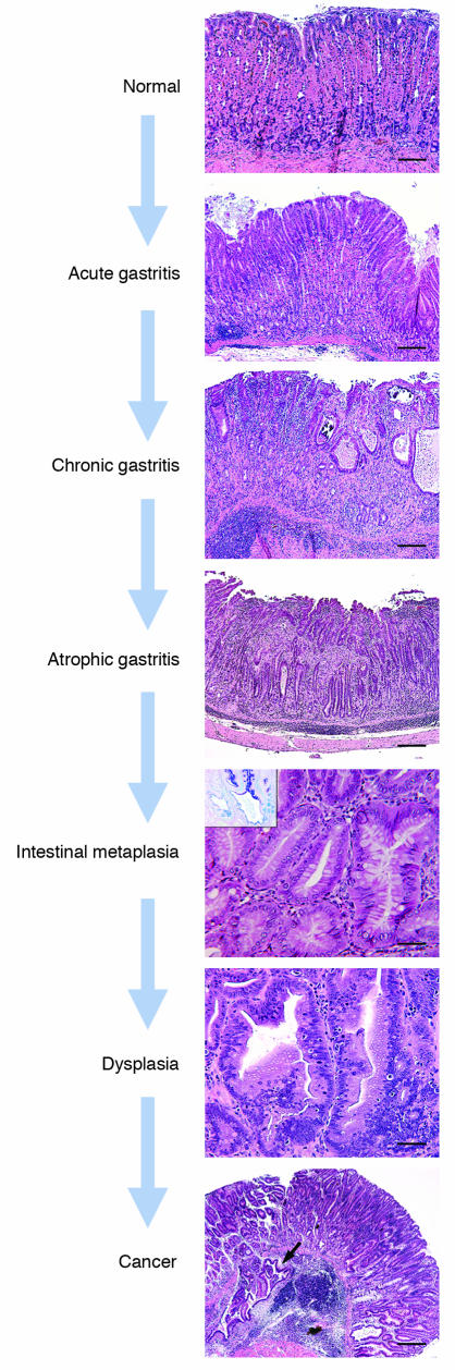
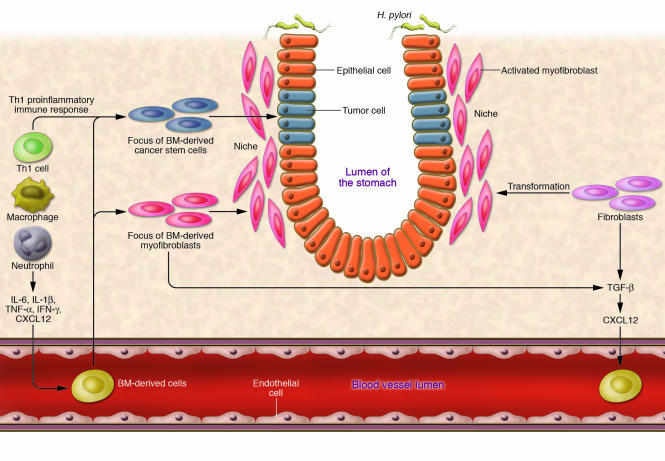
The association between chronic inflammation and cancer is now well established. This association has recently received renewed interest with the recognition that microbial pathogens can be responsible for the chronic inflammation observed in many cancers, particularly those originating in the gastrointestinal system. A prime example is Helicobacter pylori, which infects 50% of the world's population and is now known to be responsible for inducing chronic gastric inflammation that progresses to atrophy, metaplasia, dysplasia, and gastric cancer. This Review provides an overview of recent progress in elucidating the bacterial properties responsible for colonization of the stomach, persistence in the stomach, and triggering of inflammation, as well as the host factors that have a role in determining whether gastritis progresses to gastric cancer. We also discuss how the increased understanding of the relationship between inflammation and gastric cancer still leaves many questions unanswered regarding recommendations for prevention and treatment.


Figures

Figure 1
Figure 1. Diagram depicting anatomy of the stomach and histological representation of the oxyntic glands of the body of the stomach.
It is these glands, which include parietal cells, that are lost in gastric atrophy. Adapted with permission from W.B. Saunders Co. (122).
Figure 2
Figure 2. Proposed Correa pathway of pathological events in gastric adenocarcinoma.
In well-differentiated, intestinal-type gastric cancer, histopathological studies indicated that chronic H. pylori infection progresses over decades through stages of chronic gastritis, atrophy, intestinal metaplasia, dysplasia, and cancer (123). The development of cancer has been attributed to alterations in DNA caused by chronic inflammation, recruitment and engraftment of bone marrow–derived cells, an imbalance between epithelial cell proliferation and apoptosis, and, in a milieu of atrophy and achlorhydria, gastric colonization by enteric bacteria with nitrate reductase activity, which facilitates the formation of carcinogenic nitrosamines. Corpus-predominant atrophy, or the loss of specialized glandular cell types such as parietal and chief cells, appears to be the critical initiating step in the progression toward cancer. Adapted with permission from the New England Journal of Medicine (14).
Figure 3
Figure 3. H. pylori induces proinflammatory responses in epithelial cells by 2 pathways.
H. pylori–induced proinflammatory responses are dependent on the presence in H. pylori of a functional type IV secretion system (T4SS), which delivers effector molecules, such as cell wall peptidoglycan (PGN) and the protein CagA, to epithelial cells. The precise mechanisms by which the H. pylori T4SS mediates effector delivery to host cells are, however, presently unknown. (i) Recognition of H. pylori PGN by the cytosolic host defense molecule NOD1 leads to NF-κB activation. On the basis of studies with Shigella flexneri, it is likely that NOD1 activates NF-κB via caspase-recruitment domain (CARD)–CARD interactions with receptor-interacting serine-threonine kinase (RICK; also known as RIP2). Activated NF-κB complexes translocate to the nucleus, where they upregulate expression of genes encoding the proinflammatory chemokines IL-8 and CXC-chemokine ligand 2 (CXCL2; also known as MIP2) and the antimicrobial peptide human β-defensin-2 (hBD-2). (ii) CagA translocation into epithelial cells by certain H. pylori strains able to induce high levels of IL-8 is also accompanied by the induction of an inflammatory response. Tyrosine phosphorylation of EPIYA motifs on CagA triggers a signaling cascade that involves the Ras-dependent kinases ERK1 and ERK2, leading to activation of the transcription factors NF-κB and activator protein-1 (AP-1) and, ultimately, IL-8 production by host cells. Courtesy of R. Ferrero (Monash University, Clayton, Victoria, Australia).
Figure 4
Figure 4. Histological progression of Helicobacter-induced gastric cancer in a mouse model.
Normal: Histology of the body of the stomach. Acute gastritis: Infiltration of mucosal and submucosal lymphocytes with pockets of polymorphonuclear cells, accompanied by mild mucosal defects and edema. Chronic gastritis: Moderate to severe inflammation with marked epithelial defects including gland dilatation and mineralization. Atrophic gastritis: Chronic inflammation with focal fibrosis and complete loss of oxyntic parietal and chief cells. Intestinal metaplasia: Gastric epithelial metaplasia to an intestinal phenotype characterized by columnar elongation, mucous droplets occasionally forming goblet cells, and production of mixed acidic (blue, intestinal-type) and neutral (red, gastric-type) mucins as shown by pH 2.5 Alcian blue/PAS stain (inset). Dysplasia: High-grade glandular dysplasia characterized by irregular size and shape, infolding, branching and cell piling, and marked cellular and nuclear atypia. Cancer: Gastric intraepithelial neoplasia, here with intramucosal invasion (arrow), develops in H. pylori–infected wild-type B6129 mice, as well as in certain genetically engineered models (–126). Scale bars: 160 μm (first panel); 400 μm (second through fourth panels); 80 μm (fifth panel; inset, original magnification, ×400); 40 μm (sixth panel); 800 μm (seventh panel). Second panel (acute gastritis): adapted with permission from American Journal of Physiology-Gastrointestinal and Liver Physiology (124). All other panels: adapted from Cancer Research (125, 126).
Figure 5
Figure 5. BMDCs in gastric tumor initiation and progression.
H. pylori–associated chronic inflammation, driven by macrophages and Th1-polarized lymphocytes, results in increased cytokine and chemokine production that mobilizes and recruits BMDCs from the circulation. The proinflammatory milieu leads to an altered stem cell niche, characterized by increased numbers of activated myofibroblasts, which can originate from both BMDCs and local stromal populations. Some of the BMDCs may also be recruited into the progenitor zone, giving rising to epithelial metaplasia and dysplasia (i.e., cancer stem cells).

References

    1. Ries, L.A.G., et al. 2005. SEER Cancer Statistics Review, 1975–2002. National Cancer Institute. Bethesda, Maryland, USA. http://seer.cancer.gov/csr/1975_2002/.
    1. Parkin D.M., Pisani P., Ferlay J. Global cancer statistics. CA Cancer J. Clin. 1999;49:33–64. - PubMed
    1. Lauren P. The two histological main types of gastric carcinoma: diffuse and so-called intestinal-type carcinoma. An attempt at a histo-clinical classification. Acta Pathol. Microbiol. Scand. 1965;64:31–49. - PubMed
    1. Cuello C., et al. Histopathology of gastric dysplasias: correlations with gastric juice chemistry. Am. J. Surg. Pathol. 1979;3:491–500. - PubMed
    1. Philip M., Rowley D.A., Schreiber H. Inflammation as a tumor promoter in cancer induction. Semin. Cancer Biol. 2004;14:433–439. - PubMed

Publication types