Review
. 2007 Jan;117(1):70-80.
doi: 10.1172/JCI30491.

Ménétrier disease and gastrointestinal stromal tumors: hyperproliferative disorders of the stomach

Affiliations
Review

Ménétrier disease and gastrointestinal stromal tumors: hyperproliferative disorders of the stomach

Robert J Coffey et al. J Clin Invest. 2007 Jan.

Abstract

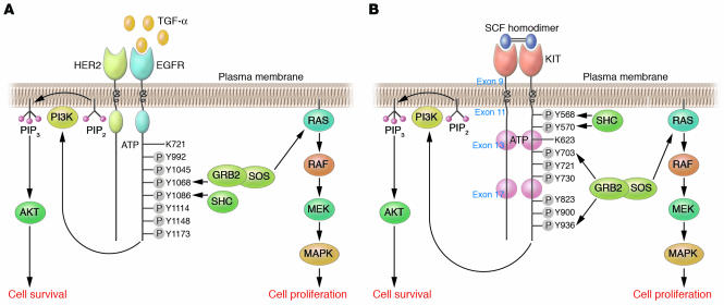
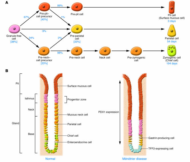
Ménétrier disease and gastrointestinal stromal tumors (GISTs) are hyperproliferative disorders of the stomach caused by dysregulated receptor tyrosine kinases (RTKs). In Ménétrier disease, overexpression of TGF-alpha, a ligand for the RTK EGFR, results in selective expansion of surface mucous cells in the body and fundus of the stomach. In GISTs, somatic mutations of the genes encoding the RTK KIT (or PDGFRA in a minority of cases) result in constitutive kinase activity and neoplastic transformation of gut pacemaker cells (interstitial cells of Cajal). On the basis of the involvement of these RTKs in the pathogenesis of these disorders, Ménétrier disease patients have been effectively treated with a blocking monoclonal antibody specific for EGFR and GIST patients with KIT and PDGFRA tyrosine kinase inhibitors.


Figures

Figure 1
Figure 1. Anatomy of the human stomach.
The inset provides more detail of the layers of the stomach wall and indicates the location of the myenteric plexus of Auerbach. Interstitial cells of Cajal (ICCs), which are specialized, spindle-shaped cells located in the gut wall that function as pacemaker cells and help regulate slow-wave peristalsis of the gut, form a network around the myenteric plexus of Auerbach and serve to regulate neural input to the smooth muscle cells of the muscularis propria. The ICC cell bodies lie in close proximity to myenteric nerves, whereas their processes extend deeply into the inner and outer layers of the muscularis, contacting individual myocytes.
Figure 2
Figure 2. Gross and microscopic view of Ménétrier disease and GIST.
(A and B) Gross (A) and microscopic (B) appearance of the stomach of a patient with Ménétrier disease (original magnification of B, ×40). (D and E) Gross (D) and microscopic (E) appearance of the stomach of a patient with a submucosal gastric GIST (original magnification of E, ×200). In D, the mucosa is stretched over the submucosal GIST with its typical central degenerative changes. (E) Typical spindle-shaped appearance of a GIST; less common are epithelioid-shaped and mixed epithelioid- and spindle-shaped GISTs. (C) TGF-α immunoreactivity in the gastric mucosa of a Ménétrier disease patient (original magnification, ×100; inset, ×300). (F) KIT immunoreactivity in a GIST (original magnification, ×200). The rulers in A and D are the same scale.
Figure 3
Figure 3. Schematic depiction of EGFR and KIT signaling.
(A) Expression of TGF-α is upregulated in the gastric mucosa of patients with Ménétrier disease. Ligand binding results in a conformational change in the ectodomain of the receptor from a tethered intramolecular state to a dimer-competent state (27). Homodimerization and/or heterodimerization with other EGFR family members results in activation of the intrinsic tyrosine kinase that phosphorylates, probably in trans, tyrosine residues (Y) in the cytoplasmic tail. Shown are the 7 autophosphorylation sites in the EGFR cytoplasmic tail. SH2-containing proteins bind these phosphorylated tyrosine residues to initiate a complex signaling cascade. GRB2 and SHC are reported to bind Y1068, Y1086, Y1148, and Y1173 (27). Activation or inhibition of EGFR usually results in coordinate regulation of MAPK and AKT, which are linked to proliferation and cell survival, respectively. (B) SCF is the only known KIT ligand. Binding of homodimeric SCF to KIT results in receptor homodimerization, activation of the intrinsic tyrosine kinase domain, and trans-phosphorylation of cytoplasmic KIT tyrosine residues. Somatic mutation of exon 9, 11, 13, or 17 in GISTs results in constitutive, ligand-independent KIT activation. Depicted are 8 tyrosine autophosphorylation sites in the KIT cytoplasmic domain. SH2-containing proteins bind these phosphorylated tyrosine residues and are then phosphorylated on tyrosine residues by KIT to initiate a complex signaling cascade. Y823 is located in the KIT activation loop; phosphorylation of this residue helps stabilize the activation loop in the “active” state and allows binding of protein substrates to the active site. Activation of KIT results in coordinate regulation of MAPK and AKT (108).
Figure 4
Figure 4. Ontogeny of cell lineages in the normal gastric body and proposed model for the pathogenesis of Ménétrier disease.
(A) In the isthmus, the stationary undifferentiated granule-free cell gives rise to pit and gland precursor cells that migrate and ultimately mature into surface mucous cells, parietal cells, and chief cells. The percentage of cells estimated to follow each differentiation pathway is indicated. The numbers in brackets indicate percentages of these progenitor cells that were found by Karam and Leblond to be proliferating; it was calculated that there was an approximately 3-day turnover time (the time required to replace the number of cells equal to the entire population) for these 4 cell types (65). Lifespans of surface mucous cells, parietal cells, and chief cells have been calculated to be 3 days, 54 days, and 194 days, respectively (65). Enteroendocrine cells also were thought by these investigators to emerge from the granule-free cell. (B) In Ménétrier disease, the progenitor compartment is expanded and repositioned at the base of the gland. We propose that this is due to increased signaling through EGFR and decreased levels of gastrin. There is expansion of the surface mucous cell compartment at the expense of parietal cells and chief cells. PDX1, pancreatic and duodenal homeobox gene 1; TFF2, trefoil factor 2.

Similar articles

Cited by

References

    1. Ménétrier P. Des polyadenomes gastriques et leur rapport avec le cancer de l’estomac. Arch. Physiol. Norm. Pathol. 1888;1:32–55; 236–262.
    1. Occena R.O., Taylor S.F., Robinson C.C., Sokol R.J. Association of cytomegalovirus with Ménétrier’s disease in childhood: report of two new cases with a review of literature. J. Pediatr. Gastroenterol. Nutr. 1993;17:217–224. - PubMed
    1. Mosnier J.F., et al. Hypertrophic gastropathy with gastric adenocarcinoma: Ménétrier’s disease and lymphocytic gastritis? Gut. 1991;32:1565–1567. - PMC - PubMed
    1. Scharschmidt B.F. The natural history of hypertrophic gastrophy (Ménétrier’s disease). Report of a case with 16 year follow-up and review of 120 cases from the literature. Am. J. Med. 1977;63:644–652. - PubMed
    1. Vandenborre K.M., et al. Hypertrophic lymphocytic gastritis with a gastric carcinoma. Eur. J. Gastroenterol. Hepatol. 1998;10:797–801. - PubMed

Publication types

MeSH terms

Substances