. 2007 Jan;117(1):153-64.
doi: 10.1172/JCI29154.

Liver heparan sulfate proteoglycans mediate clearance of triglyceride-rich lipoproteins independently of LDL receptor family members

Affiliations

Liver heparan sulfate proteoglycans mediate clearance of triglyceride-rich lipoproteins independently of LDL receptor family members

Jennifer M MacArthur et al. J Clin Invest. 2007 Jan.

Abstract

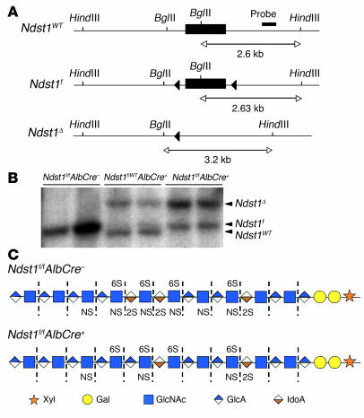
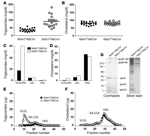
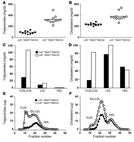
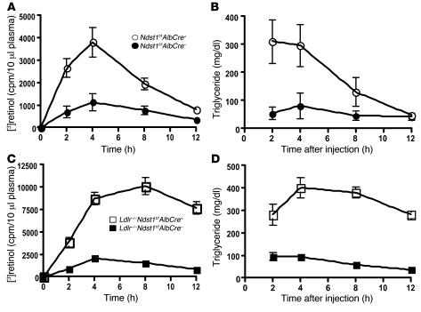
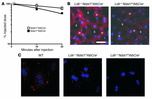
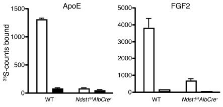
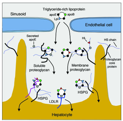
We examined the role of hepatic heparan sulfate in triglyceride-rich lipoprotein metabolism by inactivating the biosynthetic gene GlcNAc N-deacetylase/N-sulfotransferase 1 (Ndst1) in hepatocytes using the Cre-loxP system, which resulted in an approximately 50% reduction in sulfation of liver heparan sulfate. Mice were viable and healthy, but they accumulated triglyceride-rich lipoprotein particles containing apoB-100, apoB-48, apoE, and apoCI-IV. Compounding the mutation with LDL receptor deficiency caused enhanced accumulation of both cholesterol- and triglyceride-rich particles compared with mice lacking only LDL receptors, suggesting that heparan sulfate participates in the clearance of cholesterol-rich lipoproteins as well. Mutant mice synthesized VLDL normally but showed reduced plasma clearance of human VLDL and a corresponding reduction in hepatic VLDL uptake. Retinyl ester excursion studies revealed that clearance of intestinally derived lipoproteins also depended on hepatocyte heparan sulfate. These findings show that under normal physiological conditions, hepatic heparan sulfate proteoglycans play a crucial role in the clearance of both intestinally derived and hepatic lipoprotein particles.


Figures

Figure 1
Figure 1. Ndst1 conditional knockout.
(A) Gene schematic and Southern blotting strategy for mapping Ndst1 alleles. Filled triangles represent loxP recombination sites; bar indicates target sequence of Ndst1 probe; restriction sites for BglII and HindIII and expected fragment sizes are also indicated. (B) Southern blot to determine Cre-mediated recombination of Ndst1 floxed alleles. Hepatocyte genomic DNA was digested with BglII/HindIII and probed for Ndst1 alleles. The deleted allele gave a BglII/HindIII fragment at 3.2 kb, and the Ndst1f and wild-type alleles gave BglII/HindIII fragments at 2.6 kb. The Ndst1f BglII/HindIII fragment was slightly larger than 2.6 kb due to the inserted 34-bp loxP site. DNA in each lane was isolated from individual mice. Quantification of bands indicated 65%–75% recombination in AlbCre+ hepatocytes. (C) The HS chains are depicted using the indicated symbol nomenclature for the individual sugar residues. Heparinases degrade the chains to disaccharides (dashed lines), which can then be separated according the number and pattern of sulfate groups (Table 1). HS chains from the mutant contain less sulfate and iduronic acid because Ndst1 creates the preferred substrates for epimerization and O-sulfation. The residual sulfation presumably arises from incomplete inactivation of Ndst1 and the expression of Ndst2.
Figure 2
Figure 2. Hypertriglyceridemia in Ndst1-deficient mice.
Total triglycerides (A) and total cholesterol (B) were measured in plasma samples (n = 20 per genotype, mixed males and females, horizontal bars indicate mean values). Triglyceride (C) and cholesterol (D) content of lipoproteins fractionated by preparative density ultracentrifugation was determined. Each value is from 1 set of pooled plasma derived from 10 mice. Lipoproteins were also analyzed by FPLC gel filtration, and the amount of triglyceride (E) and cholesterol (F) was determined in control mice (filled circles) and Ndst1f/fAlbCre+ mice (open circles). Plasma was pooled from 9 mice of each genotype. The elution positions of human VLDL, IDL/LDL, and HDL are indicated. (G) Samples of purified lipoproteins (d < 1.019 g/ml) were analyzed by gradient SDS-PAGE, and the individual apolipoproteins were visualized by Coomassie blue or silver staining. The location of apoB-48, apoB-100, apoE, and apoAI was determined by Western blotting (data not shown). The location of apoCs and serum albumin was deduced by Mr values.
Figure 3
Figure 3. Cholesterol and triglyceride particles accumulated in Ldlr–/–Ndst1f/fAlbCre+ mice.
Total plasma triglyceride (A) and cholesterol (B) levels were determined for Ldlr–/–Ndst1f/fAlbCre mice (filled circles) and Ldlr–/–Ndst1f/fAlbCre+ mice (open circles). Points represent individual animals; n = 11 per genotype; horizontal bars indicate mean values. All mice (8–10 weeks old) were fed normal chow and fasted overnight prior to analysis. Double mutants showed significant accumulation of both triglycerides and cholesterol. Triglyceride (C) and cholesterol (D) content of lipoproteins fractionated according to density by preparative ultracentrifugation. Each value is from 1 set of pooled plasma derived from 10 mice. Lipoproteins were analyzed by FPLC gel filtration, and the amount of triglyceride (E) and cholesterol (F) was determined in Ldlr–/–Ndst1f/fAlbCre mice (filled circles) and Ldlr–/–Ndst1f/fAlbCre+ mice (open circles). Plasma was pooled from 10 mice of each genotype. The elution positions of human VLDL, IDL/LDL, and HDL are indicated.
Figure 4
Figure 4. Slowed clearance of postprandial lipoproteins.
Retinyl ester excursions and plasma triglyceride levels were measured at the times indicated in 8-week-old Ndst1f/fAlbCre+ mice (n = 9) compared with wild-type siblings (n = 5) (A and B) and Ldlr–/–Ndst1f/fAlbCre+ mice (n = 7) compared with Ldlr–/–Ndst1f/fAlbCre siblings (n = 4) (C and D). Overnight-fasted animals of the indicated genotypes were given 200 μl of corn oil containing 5.4 μCi [3H]retinol by gavage. Blood samples were taken at the indicated times, and radioactivity remaining in 10 μl of serum was determined by scintillation counting in triplicate. The values are expressed as mean ± SD.
Figure 5
Figure 5. Decreased plasma clearance and liver uptake of VLDL in Ndst1-deficient mice.
(A) Plasma clearance of human VLDL apoB-100 was measured by ELISA using human apoB-100–specific monoclonal antibody MB47 (see Methods). Control mice (filled squares; t1/2 = 23.2 ± 1.4 minutes; n = 6) and Ndst1f/fAlbCre+ mice (open squares; t1/2 = 46.6 ± 5.3 minutes; n = 6). The difference in t1/2 between the genotypes was significant (P = 0.0016), and the difference between the 10- and 20-minute time points for the mutant was also significant (P = 0.0133). Results were verified in a separate experiment. (B) Deconvolution microscopy showing uptake of DiI-VLDL (red) in the liver of Ldlr–/–Ndst1f/fAlbCre and Ldlr–/–Ndst1f/fAlbCre+ mice. DiI-VLDL (100 μg) was injected by tail vein, and livers were fixed 20 minutes later. Sections of 20 μm were DAPI-stained for nuclei (blue). H, hepatocyte cords; S, sinusoids. Scale bar: 26 μm; magnification, ×40. (C) Hepatocytes were isolated from mice of the indicated genotypes and incubated with DiI-VLDL (5 μg/ml). Images were obtained by deconvolution microscopy.
Figure 6
Figure 6. Altered binding of mutant HS to apoE and FGF2.
Radiolabeled HS was isolated from cultured primary hepatocytes from Ndst1f/fAlbCre+ mice or wild-type controls grown in the presence of 35SO4 (see Methods). Samples (104 cpm) were incubated in physiologic saline with 10 μg recombinant apoE (lipid-free) or FGF2 in the absence (white bars) and presence (black bars) of 100 μg/ml of heparin, and bound material was collected by membrane filtration. Each experiment was done in triplicate, and the average values are shown.
Figure 7
Figure 7. Model of possible roles for hepatic HS in TRL clearance.
Hepatocytes and endothelial cells produce membrane-bound HSPGs and secrete proteoglycans into the space of Disse. After lipolytic processing of lipoproteins in the circulation by Lpl (blue triangles), apoE-enriched (black circles) remnant lipoproteins enter the space of Disse through fenestrations in the endothelium. Remnant lipoproteins are thought to be sequestered near the hepatocyte cell surface via apoE-HS binding or lipase-HS bridging on secreted HSPGs. Lipoproteins are further processed in the space of Disse by transfer of soluble apoE (gray circles) and by HL (red triangles) bound via HS. apoE, HL, and Lpl can potentially serve as ligands of TRLs. Endocytosis of lipoprotein particles occurs via LDLR (blue) or LRP (purple) in association with HSPGs or independently by proteoglycans. Adapted with permission from Journal of Lipid Research (8) and Arteriosclerosis, Thrombosis, and Vascular Biology (86).

Comment in

Similar articles

Cited by

References

    1. Conrad, H.E. 1998. Heparin-binding proteins. Academic Press. San Diego, California, USA. 527 pp.
    1. Bernfield M., et al. Functions of cell surface heparan sulfate proteoglycans. Annu. Rev. Biochem. 1999;68:729–777. - PubMed
    1. Esko J.D., Selleck S.B. Order out of chaos: assembly of ligand binding sites in heparan sulfate. Annu. Rev. Biochem. 2002;71:435–471. - PubMed
    1. Williams K.J., Fuki I.V. Cell-surface heparan sulfate proteoglycans: dynamic molecules mediating ligand catabolism. Curr. Opin. Lipidol. 1997;8:253–262. - PubMed
    1. Belting M. Heparan sulfate proteoglycan as a plasma membrane carrier. Trends Biochem. Sci. 2003;28:145–151. - PubMed

Publication types

MeSH terms