. 2007 Jan;117(1):165-74.
doi: 10.1172/JCI29415.

Disruption of LDL but not VLDL clearance in autosomal recessive hypercholesterolemia

Affiliations

Disruption of LDL but not VLDL clearance in autosomal recessive hypercholesterolemia

Christopher Jones et al. J Clin Invest. 2007 Jan.

Abstract

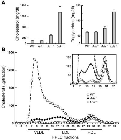
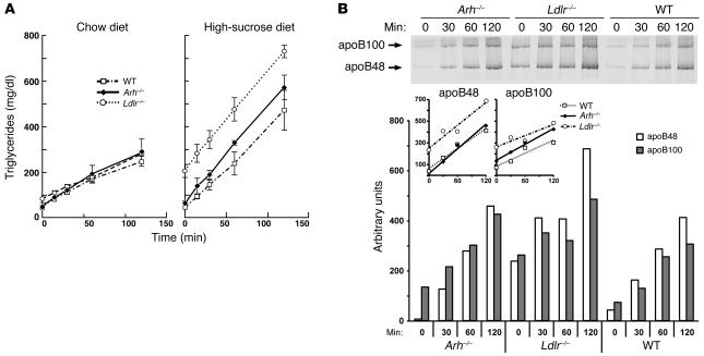
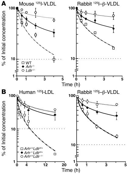
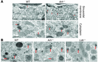
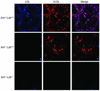
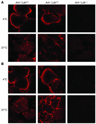
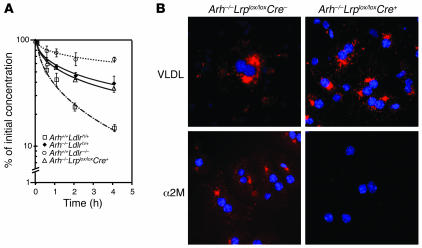
Genetic defects in LDL clearance result in severe hypercholesterolemia and premature atherosclerosis. Mutations in the LDL receptor (LDLR) cause familial hypercholesterolemia (FH), the most severe form of genetic hypercholesterolemia. A phenocopy of FH, autosomal recessive hypercholesterolemia (ARH), is due to mutations in an adaptor protein involved in LDLR internalization. Despite comparable reductions in LDL clearance rates, plasma LDL levels are substantially lower in ARH than in FH. To determine the metabolic basis for this difference, we examined the synthesis and catabolism of VLDL in murine models of FH (Ldlr(-/-)) and ARH (Arh(-/-)). The hyperlipidemic response to a high-sucrose diet was greatly attenuated in Arh(-/-) mice compared with Ldlr(-/-) mice despite similar rates of VLDL secretion. The rate of VLDL clearance was significantly higher in Arh(-/-) mice than in Ldlr(-/-) mice, suggesting that LDLR-dependent uptake of VLDL is maintained in the absence of ARH. Consistent with these findings, hepatocytes from Arh(-/-) mice (but not Ldlr(-/-) mice) internalized beta-migrating VLDL (beta-VLDL). These results demonstrate that ARH is not required for LDLR-dependent uptake of VLDL by the liver. The preservation of VLDL remnant clearance attenuates the phenotype of ARH and likely contributes to greater responsiveness to statins in ARH compared with FH.


Figures

Figure 1
Figure 1. Plasma cholesterol and triglyceride levels (A) and lipoprotein cholesterol levels (B) in sucrose-fed wild-type, Ldlr–/–, Arh+/–, and Arh–/– mice.
Male Arh–/–, Arh+/–, and wild-type littermates, ages 16–18 weeks, and age-matched Ldlr–/– mice (n = 6 per genotype) were fed a high-sucrose diet for 6 weeks. Mice were sacrificed and blood was drawn from the inferior vena cava. Individual plasma cholesterol and triglyceride levels (A) were measured as described in Methods, and the means ± SEM are given. (B) Aliquots of plasma from the animals in each group were pooled and size fractionated by FPLC. The cholesterol content of each fraction was measured as described (39).
Figure 2
Figure 2. VLDL production rates of Arh–/–, Ldlr–/–, and wild-type mice on normal chow and high-sucrose diets.
(A) Male Arh–/– (diamonds), Ldlr–/– (circles), and wild-type (squares) mice (14–16 weeks of age) were injected with 300 mg/kg Triton WR-1339 (15% w/v in 0.9% NaCl). Prior to injection, the mice were maintained on a normal chow diet (left) or a high-sucrose diet (right) for 6 weeks. Blood was sampled from the retro-orbital plexus after a 6-hour fast at the time points indicated, and the triglycerides were measured enzymatically as described in Methods. VLDL production rates were calculated from the slope of a line determined using least squares regression. (B) apoB production in Arh–/–, Ldlr–/–, and wild-type mice. Male Arh–/– (diamonds), Ldlr–/– (circles), and wild-type (squares) mice (n = 4), 18–22 weeks of age, on a chow diet were injected with 300 mg/kg Triton WR-1339 (15% w/v in 0.9% NaCl) after a 6-hour fast. Blood was sampled from the retro-orbital plexus at the times indicated. Plasma aliquots from individual mice were pooled, and VLDL was separated as described in Methods. The resulting apolipoproteins were separated by SDS-PAGE and stained with colloidal Coomassie blue. Scanning densitometry was used to determine the relative amounts of apoB48 and apoB100 in each sample.
Figure 3
Figure 3. Clearance of circulating VLDL but not LDL is partially preserved in Arh–/– mice.
(A) Clearance of mouse 125I-VLDL and rabbit 125I–β-VLDL in Arh–/–, Ldlr–/–, and wild-type mice. Four wild-type (squares), Arh–/– (diamonds), and Ldlr–/– (circles) 12- to 14-week-old female mice were injected with mouse 125I-VLDL (125 cpm/ng protein) (left) or rabbit 125I–β-VLDL (288 cpm/ng protein) (right). Blood samples were collected by retro-orbital puncture at the indicated times, and the plasma content of isopropanol-precipitable 125I-radioactivity was measured. Radioactivity remaining in the plasma was plotted as a percentage of the activity present 2 minutes after injection of the labeled ligand. Before the experiment, the mice were fasted for 6 hours and anesthetized with sodium pentobarbital (80 mg/kg intraperitoneal). (B) Female Arh+/+Ldlrh/+ (squares), Arh–/–Ldlrh/+ (diamonds), and Ldlr–/– (circles) mice (n = 4 per genotype), aged 12–14 weeks, were fasted for 4 hours, anesthetized with sodium pentobarbital (80 mg/kg of intraperitoneal), and injected with 15 μg 125I-human LDL (125 cpm/ng protein) (left) or 15 μg 125I-rabbit β-VLDL (189 cpm/ng protein) (right). Blood samples were collected and processed as described above.
Figure 4
Figure 4. Localization of LDL-gold (A) and β-VLDL–gold (B) in Ldlrh/h, Ldlrh/hArh–/–, and Ldlr–/– mice.
Mice were injected via the tail vein with 50 μg of colloidal gold–labeled human LDL or rabbit β-VLDL. After 2 hours, mice were sacrificed. Livers were perfused, fixed, and then processed for electron microscopy as described in Methods. Solid arrows indicate gold particles associated with villi on the sinusoidal membrane, and open arrows indicate endosomes. Scale bars: 0.5 μm.
Figure 5
Figure 5. PMCA-O LDL and DiI β-VLDL uptake by primary hepatocytes from Arh+/+Ldlrh/h, Arh–/–Ldlrh/h, and Ldlr–/– mice.
On day 0, primary hepatocytes were isolated from Arh+/+Ldlrh/h, Arh–/–Ldlrh/h, and Ldlr–/– mice and incubated overnight in DMEM containing 5% lipoprotein-deficient serum. The next morning, the cells were incubated at 37°C in fresh media containing 25 μg PMCA-O LDL and 25 μg DiI β-VLDL for 5 hours. The cells were washed, fixed, and mounted as described in Methods. Images were procured by deconvolution microscopy. Original magnification, ×84.
Figure 6
Figure 6. Internalization of IgG-C7 and IgG-C7:protein A complexes in primary hepatocytes.
Primary hepatocytes were isolated from Arh+/+Ldlrh/h, Arh–/–Ldlrh/h, and Arh+/+Ldlr–/– mice and incubated for 1 hour at 4°C with either (A) IgG-C7 or (B) IgG-C7 complexed with protein A. Cells were then warmed to 37°C for 2 hours. IgG-C7 was detected by indirect immunofluorescence confocal microscopy. Original magnification, ×130.
Figure 7
Figure 7. Deletion of LRP does not reduce β-VLDL clearance in Arh–/– mice.
(A) Mice (n = 3 per group) were fasted for 4 hours, anesthetized with sodium pentobarbital, and injected with 125I-labeled rabbit β-VLDL (15 μg) via the external jugular vein. Venous blood was collected from the retro-orbital plexus at the indicated times, and the plasma content of isopropanol-precipitable 125I-radioactivity was measured. Radioactivity remaining in the plasma was plotted as a percentage of the activity present 2 minutes after injection of the labeled ligand. The experiment was repeated, and similar results were obtained. (B) Primary hepatocytes were isolated from Arh–/–Lrplox/loxCre and Arh–/–Lrplox/loxCre+ mice and incubated overnight in DMEM containing 5% lipoprotein-deficient serum. The next morning, the cells were incubated with 15 μg/ml DiI β-VLDL or 10 μg/ml methylamine-activated α2-macroglobulin (α2M) (51) for 30 minutes. The cells were washed, fixed, and mounted as described in Methods. Images were taken by deconvolution microscopy. Original magnification, ×84.

Comment in

Similar articles

Cited by

References

    1. Goldstein, J., Hobbs, H., and Brown, M. 2001. Familial hypercholesterolemia. In The metabolic and molecular bases of inherited disease. C. Scriver, A. Beaudet, W. Sly, and D. Valle, editors. McGraw-Hill. New York, New York, USA. 2863–2913.
    1. Khachadurian A.K., Uthman S.M. Experiences with the homozygous cases of familial hypercholesterolemia. A report of 52 patients. Nutr. Metab. 1973;15:132–140. - PubMed
    1. Norman D., et al. Characterization of a novel cellular defect in patients with phenotypic homozygous familial hypercholesterolemia. J. Clin. Invest. 1999;104:619–628. - PMC - PubMed
    1. Zuliani G., et al. Severe hypercholesterolaemia: unusual inheritance in an Italian pedigree. Eur. J. Clin. Invest. 1995;25:322–331. - PubMed
    1. Harada-Shiba M., et al. Siblings with normal LDL receptor activity and severe hypercholesterolemia. Arterioscler. Thromb. 1992;12:1071–1078. - PubMed

Publication types