. 2007 Jan;117(1):195-205.
doi: 10.1172/JCI29950.

Ly-6Chi monocytes dominate hypercholesterolemia-associated monocytosis and give rise to macrophages in atheromata

Affiliations

Ly-6Chi monocytes dominate hypercholesterolemia-associated monocytosis and give rise to macrophages in atheromata

Filip K Swirski et al. J Clin Invest. 2007 Jan.

Abstract

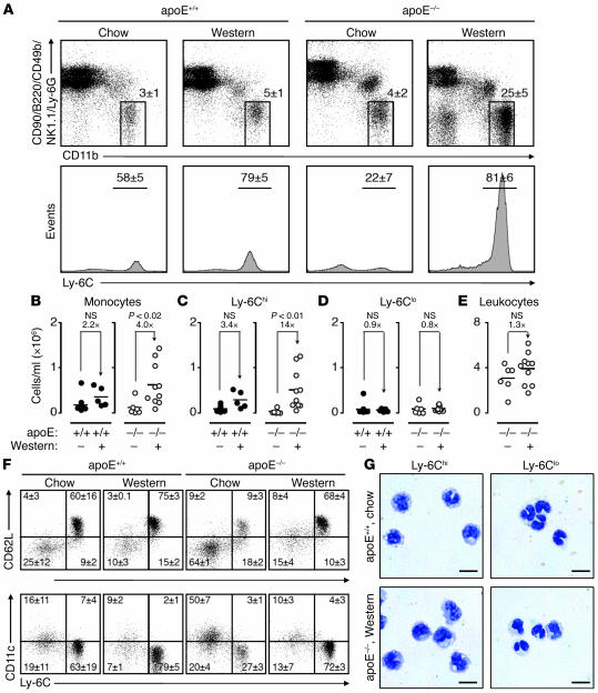
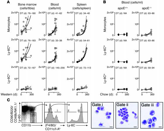
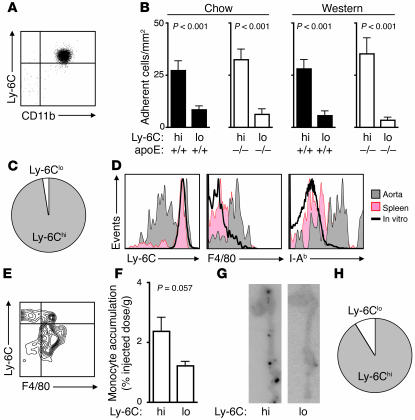
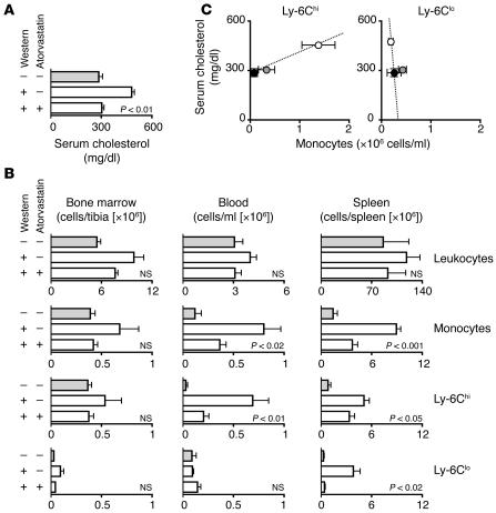
Macrophage accumulation participates decisively in the development and exacerbation of atherosclerosis. Circulating monocytes, the precursors of macrophages, display heterogeneity in mice and humans, but their relative contribution to atherogenesis remains unknown. We report here that the Ly-6C(hi) monocyte subset increased dramatically in hypercholesterolemic apoE-deficient mice consuming a high-fat diet, with the number of Ly-6C(hi) cells doubling in the blood every month. Ly-6C(hi) monocytes adhered to activated endothelium, infiltrated lesions, and became lesional macrophages. Hypercholesterolemia-associated monocytosis (HAM) developed from increased survival, continued cell proliferation, and impaired Ly-6C(hi) to Ly-6C(lo) conversion and subsided upon statin-induced cholesterol reduction. Conversely, the number of Ly-6C(lo) cells remained unaffected. Thus, we believe that Ly-6C(hi) monocytes represent a newly recognized component of the inflammatory response in experimental atherosclerosis.


Figures

Figure 1
Figure 1. Hypercholesterolemia induces peripheral blood Ly-6Chi monocytosis.
(A) Mononuclear cells from blood of apoE+/+ and apoE–/– mice consuming either chow or Western diet were stained with anti-CD11b, -CD90, -B220, -CD49b, -NK1.1, –Ly-6G, and –Ly-6C mAbs. Living cells were gated to determine presence and percentage of CD11bhiCD90loB220loCD49bloNK1.1loLy-6Glo monocytes (top row) and further divided into Ly-6Chi and Ly-6Clo subsets (bottom row). Representative dot plots and histograms from individual mice are depicted. Percentages of cells are shown as mean ± SEM. (B) Total blood monocytes in apoE+/+ and apoE–/– mice consuming either Western diet (+) or chow (–). (C) Total blood Ly-6Chi monocytes. (D) Total blood Ly-6Clo monocytes. (E) Total peripheral blood leukocytes. (F) Representative dot plots depicting expression of CD62L and CD11c among Ly-6Chi and Ly-6Clo monocytes. Percentages of cells in each quadrant are shown as mean ± SEM. (G) Representative cytospin preparations of purified blood Ly-6Chi and Ly-6Clo monocytes in apoE+/+ mice on chow and apoE–/– mice on Western diet. Scale bar: 10 μm. Student’s t test was used. Results are representative of 8 independent experiments with 5–14 mice per group.
Figure 2
Figure 2. Peripheral blood monocytosis develops over the course of 250 days on an atherogenic diet.
(A) Number of total monocytes and Ly-6Chi and Ly-6Clo subtypes in bone marrow, blood, and spleens of apoE–/– mice at various days of Western diet. Statistical analysis was based on an exponential growth curve and known cell numbers on day 0. Curve fit (solid line) and 95% confidence intervals (dashed lines) are shown. Doubling time (DT) of cell number is shown. (B) The same analysis was conducted with peripheral blood from apoE+/+ and apoE–/– mice that remained on chow diet. Doubling time of cell number in days is shown. (C) Splenic CD11bhiCD90loB220loCD49bloNK1.1loLy-6Glo cells were divided into F4/80hiCD11chiI-Ab–high macrophages/dendritic cells (gate i) and F4/80loCD11cloI-Ab–low monocytes, which were further divided into Ly-6Clo (gate ii) and Ly-6Chi (gate iii) subsets. These 3 subsets were isolated and stained with HEMA 3 for microscopic analysis. Scale bar: 10 μm. Results are pooled from 8 independent experiments.
Figure 3
Figure 3. Ly-6Chi monocytosis results from increased survival, continued proliferation, and impaired Ly-6Chi to Ly-6Clo monocyte conversion.
(A) Ly-6Chi monocytes from the spleens of apoE–/– mice were placed into culture with medium alone or medium supplemented with 100 μg/ml AcLDL or 50 μg/ml M-CSF. The percentage of cells alive 24 hours later was calculated based on the ratio of retrieved and input cell numbers. **P < 0.01, *P < 0.05 versus medium alone (1-way ANOVA with Tukey’s multiple comparison test). (B) apoE+/+ and apoE–/– mice on chow and Western diet received 3 i.p. injections of BrdU on 3 consecutive days. Cells from bone marrow, blood, and spleen were collected 1 day after the last injection and labeled with annexin V or anti-BrdU mAb. Results are shown for gated CD11bhiCD90loB220loCD49bloNK1.1loLy-6Glo monocytes as identified in Figure 1. Statistical analyses were performed using Student’s t test. (C) Representative dot plots depicting annexin V staining and BrdU incorporation in splenic CD11bhiCD90loB220loCD49bloNK1.1loLy-6Glo monocytes from apoE+/+ and apoE–/– mice on chow and Western diet. (D) apoE–/– mice on chow and Western diet received clodronate liposomes on day 0. Representative contour plots depict Ly-6C versus F4/80/I-Ab/CD11c phenotype among splenic CD11bhiCD90loB220loCD49bloNK1.1loLy-6Glo monocytes on days 1 and 5 or in age-matched untreated mice. (E) Percent of splenic Ly-6Clo monocytes recovered after clodronate liposome injection in apoE–/– mice on chow and Western diet compared with absolute number of cells in age-matched untreated mice. Shown are 1 of 2–3 independent experiments.
Figure 4
Figure 4. Atherosclerotic lesions contain Ly-6Chi monocytes.
(A) Aortas from apoE+/+ and apoE–/– mice on chow and Western diet were digested with a protease cocktail. Cells were dispersed and stained with anti-CD11b, -CD90, -B220, -CD49b, -NK1.1, –Ly-6G, -F4/80, –I-Ab, -CD11c, and –Ly-6C mAb. Percent (mean ± SEM) are shown for each quadrant. (B) Number of retrieved Ly-6Chi and Ly-6Clo monocytes per aorta in apoE+/+ and apoE–/– mice on chow and Western diet. Results are pooled from 5 independent experiments with 2–5 mice per group. Mean and SEM are shown. #P < 0.001 versus all other groups (1-way ANOVA with Tukey’s multiple comparison test). (C) Immunohistochemistry depicts the intima at the aortic root of a representative apoE–/– mouse on Western diet and an apoE+/+ mouse on chow. Sections stained with anti-CD31, –Ly-6C, and –Mac-3 mAbs are shown. Original magnification, ×400.
Figure 5
Figure 5. Ly-6Chi monocytes adhere preferentially to TNF-α–activated endothelium, accumulate in lesions, and differentiate to macrophages in vivo.
(A) Purified Ly-6Chi monocyte phenotype after 24 hours in culture. (B) Adherence of blood monocytes on TNF-α–treated MHECs under laminar flow conditions. Monocyte subsets were isolated from the blood of apoE+/+ and apoE–/– mice on chow and Western diet. (C) Relative proportion of blood monocytes expected to adhere to activated endothelium, based on the capacity of each subset to adhere and their average abundance in peripheral blood of apoE–/– mice on Western diet for 25 weeks. (D) Ly-6C, F4/80, and I-Ab expression of CD45.2+ Ly-6Chi monocytes retrieved from aortas and spleens 24 hours after transfer into CD45.1+ mice (both donor and recipient apoE–/– mice consuming a Western diet). Monocytes cultured in vitro were also analyzed. (E) F4/80 and Ly-6C coexpression on CD45.2+ donor cells retrieved from aortas. (F) In vivo aortic accumulation of [111In]oxine-labeled Ly-6Chi and Ly-6Clo monocytes 24 hours after adoptive transfer in apoE–/– mice on Western diet. (G) Phosphorimager plates depicting relative distribution of signal in aortas of apoE–/– mice that received equal numbers of Ly-6Chi apoE–/– or Ly-6Clo apoE–/– monocytes. (H) Relative proportion of Ly-6Chi and Ly-6Clo monocytes expected to accumulate in atherosclerotic aortas, based on the capacity of each subset for aortic accumulation and their average abundance in peripheral blood of apoE–/– mice on Western diet for 25 weeks. Shown are 1 of 2–3 independent experiments. Student’s t test was used.
Figure 6
Figure 6. Statin treatment limits monocytosis.
apoE–/– mice consumed Western diet supplemented or not with atorvastatin for 25 weeks. A control group of apoE–/– mice received regular chow. (A) Serum cholesterol after 25 weeks of diet. (B) Number of leukocytes, monocytes, and Ly-6Chi and Ly-6Clo subtypes in bone marrow, blood, and spleen. (C) Association between serum cholesterol and number of circulating Ly-6Chi or Ly-6Clo monocytes after 25 weeks of diet. Mean ± SEM are shown for apoE–/– mice on chow (filled circles), Western diet (open circles), and Western diet supplemented with atorvastatin (gray circles). Results are pooled from 9 independent experiments (n = 3–14 per group). Student’s t test was used.

Comment in

References

    1. Hansson G.K., Libby P. The immune response in atherosclerosis: a double-edged sword. Nat. Rev. Immunol. 2006;6:508–519. - PubMed
    1. Libby P. Inflammation in atherosclerosis. Nature. 2002;420:868–874. - PubMed
    1. Ridker P.M., Hennekens C.H., Buring J.E., Rifai N. C-reactive protein and other markers of inflammation in the prediction of cardiovascular disease in women. N. Engl. J. Med. 2000;342:836–843. - PubMed
    1. Peter K., et al. Circulating vascular cell adhesion molecule-1 correlates with the extent of human atherosclerosis in contrast to circulating intercellular adhesion molecule-1, E-selectin, P-selectin, and thrombomodulin. Arterioscler. Thromb. Vasc. Biol. 1997;17:505–512. - PubMed
    1. Barron H.V., Cannon C.P., Murphy S.A., Braunwald E., Gibson C.M. Association between white blood cell count, epicardial blood flow, myocardial perfusion, and clinical outcomes in the setting of acute myocardial infarction: a thrombolysis in myocardial infarction 10 substudy. Circulation. 2000;102:2329–2334. - PubMed

Publication types