. 2007 Jan;117(1):258-69.
doi: 10.1172/JCI29159.

Myd88-dependent positioning of Ptgs2-expressing stromal cells maintains colonic epithelial proliferation during injury

Affiliations

Myd88-dependent positioning of Ptgs2-expressing stromal cells maintains colonic epithelial proliferation during injury

Sarah L Brown et al. J Clin Invest. 2007 Jan.

Abstract

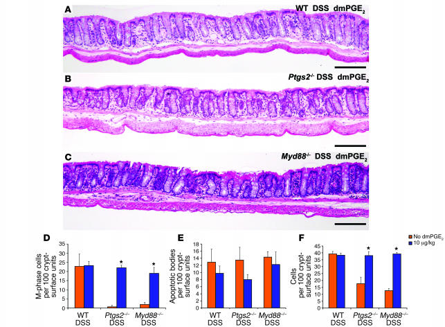
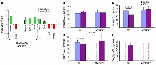
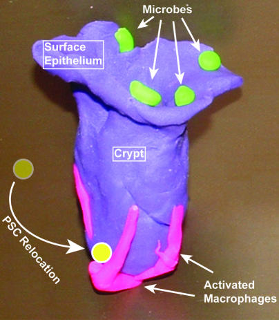
We identified cellular and molecular mechanisms within the stem cell niche that control the activity of colonic epithelial progenitors (ColEPs) during injury. Here, we show that while WT mice maintained ColEP proliferation in the rectum following injury with dextran sodium sulfate, similarly treated Myd88(-/-) (TLR signaling-deficient) and prostaglandin-endoperoxide synthase 2(-/-) (Ptgs2(-/-)) mice exhibited a profound inhibition of epithelial proliferation and cellular organization within rectal crypts. Exogenous addition of 16,16-dimethyl PGE(2) (dmPGE(2)) rescued the effects of this injury in both knockout mouse strains, indicating that Myd88 signaling is upstream of Ptgs2 and PGE(2). In WT and Myd88(-/-) mice, Ptgs2 was expressed in scattered mesenchymal cells. Surprisingly, Ptgs2 expression was not regulated by injury. Rather, in WT mice, the combination of injury and Myd88 signaling led to the repositioning of a subset of the Ptgs2-expressing stromal cells from the mesenchyme surrounding the middle and upper crypts to an area surrounding the crypt base adjacent to ColEPs. These findings demonstrate that Myd88 and prostaglandin signaling pathways interact to preserve epithelial proliferation during injury using what we believe to be a previously undescribed mechanism requiring proper cellular mobilization within the crypt niche.


Figures

Figure 1
Figure 1. Myd88–/– and Ptgs2–/– mouse rectums were susceptible to DSS-induced injury.
(A) Colon whole mount from an adult WT B6 mouse treated with 2.5% DSS for 1 week. The DSS-induced lesions (top) and the corresponding anatomic locations (bottom) are indicated. All strains analyzed in this study showed multiple small (<0.5 mm in diameter) ulcers with heaped borders in the descending colon and a confluent ulcer that extended from the ano-rectal junction proximally (0.5–1.0 cm). The intervening area in the remaining rectum (yellow boxed area) was spared of ulcerative injury. (BG) H&E-stained sections of the rectums from adult B6 WT (B and C), Myd88–/– (D and E), and Ptgs2–/– (F and G) mice. (B, D, and F) Untreated mice. (C, E, and G) Mice treated for 7 days with 2.5% DSS in the drinking water. The rectums from WT DSS-treated mice were indistinguishable from those of untreated WT mice. However, DSS-treated Myd88–/– and Ptgs2–/– mice both showed a similar pattern of crypt area loss manifested by alterations in crypt morphology (including angulation and dilation of the base; arrows) as compared with untreated controls. Scale bars: 1 cm (A), 100 μm (BG).
Figure 2
Figure 2. Goblet cell reorganization and loss of epithelial proliferation in the rectum of DSS-treated Ptgs2–/– and Myd88–/– mice.
(AF) Sections of a rectal crypt-surface unit from WT (A and B), Myd88–/– (C and D), and Ptgs2–/– (E and F) mice. (A, C, and E) Untreated mice. (B, D, and F) DSS-treated mice. Sections were stained with PAS/AB to identify goblet cells (left) or with goat anti-BrdU, Alexa-Fluor 594–labeled donkey anti-goat Ig (red), and bis-benzimide (blue nuclear stain) to identify cells in S-phase (right). Scale bars: 20 μm. The crypt epithelial-mesenchymal (dotted lines) and the epithelial crypt-surface junctions (dashed lines) are indicated. Quantification of goblet cells per crypt (G), crypt cell census (H), crypt height (I), epithelial apoptosis (J), and epithelial proliferation (K). Mean values ± SEM were plotted for each group. An asterisk indicates a value that is statistically significantly different from the corresponding untreated control (*P < 0.001; Student’s t test). Both DSS-treated Myd88–/– and Ptgs2–/– mice showed a statistically significant decrease in epithelial proliferation and crypt cell census as compared with their corresponding untreated controls.
Figure 3
Figure 3. There was no statistically significant inflammatory infiltrate in the rectum of DSS-treated mice.
Quantification of lymphocytes (B220 and CD3ε), neutrophils and eosinophils (Gr-1), and macrophages (F4/80) per crypt-surface unit. Mean values ± SEM were plotted for each group. No statistically significant differences were observed when comparing any of the DSS-treated mice with untreated mice (Student’s t test; P < 0.05).
Figure 4
Figure 4. Exogenous dmPGE2 rescued the rectal phenotype of DSS-treated Ptgs2–/– and Myd88–/– mice.
(AC) H&E-stained sections of rectums from (A) WT, (B) Ptgs2–/–, and (C) Myd88–/– mice that were concurrently treated with DSS and 10 μg/kg dmPGE2. In all cases, the low-power view of the rectum was similar to that of WT DSS-treated mice in the absence of exogenous dmPGE2 (Figure 1C). Scale bars: 100 μm. (DF) Quantification of (D) epithelial proliferation, (E) epithelial apoptosis, and (F) crypt cell census. Mean values ± SEM were plotted for each group. An asterisk indicates a value that is statistically significantly different from the corresponding control that did not receive dmPGE2 (*P < 0.001; Student’s t test). This dosage of dmPGE2 did not affect the rectum of WT DSS-treated mice and rescued the rectal phenotype of DSS-treated Ptgs2–/– and Myd88–/– mice.
Figure 5
Figure 5. Ptgs2 transcripts expressed in the lamina propria mesenchyme were not elevated in response to injury.
(A) qRT-PCR analysis of LCM-procured mRNAs from the rectal mucosal mesenchyme versus the overlying epithelium. A positive value (green) indicated transcript enrichment in the mesenchyme, and a negative value (red) indicated enrichment in the epithelium. The SEM of the fold difference for each gene was calculated from the averages of each experimental group analyzed (WT and Myd88–/– mice with and without DSS treatment). Dissection controls included vimentin, Pecam1, CD11c, and F4/80 for the mesenchyme and E-cadherin (E-cad) for the epithelium. Ptgs2, Iigp1, Indo, and Reg3g were identified in our prior microarray screens for transcripts elevated with DSS injury (boxed region). Ptgs2 and Iigp1 were enriched in the mesenchyme, while Indo and Reg3g were enriched in the epithelium. (BE) Shown are mean ± SD of ΔCTs for Ptgs2 (B), Indo (C), Iigp1 (D), and Reg3g (E) as measured from the compartment where the transcript was enriched. A smaller ΔCT indicates greater expression. The relative transcript levels for Ptgs2 were not significantly different between DSS-treated and untreated controls for either WT or Myd88–/– rectal mesenchyme. Both Iigp1 and Indo were significantly enriched in DSS-treated WT mice (versus untreated). Signal for Reg3g was only detected in the WT DSS-treated sample.
Figure 6
Figure 6. PSCs are CD44+ stromal cells.
(AI) Double-labeled, immunofluorescence-stained rectal sections from a WT DSS-treated mouse. (AC) Sections stained with Zenon Alexa Fluor 594–labeled anti-Ptgs2 Ig (Invitrogen) (PSCs, red), FITC-labeled anti-CD44 Ig (green), and bis-benzimide (blue). All PSCs show membrane staining for CD44. Arrowheads denote CD44+ epithelial cells in the crypt base. (A) Ptgs2, (B) CD44, and (C) merged image of A and B. (DF) Two crypt bases from a section stained with Zenon Alexa Fluor 594–labeled anti-Ptgs2 Ig (PSCs, red) and FITC-labeled anti-CD45 Ig (leukocytes, green). (D) Ptgs2, (E) CD45, and (F) merged images (upper panel shows a CD45 PSC, and lower panel shows a weakly CD45+ PSC). The arrowhead denotes the position of a CD45+ intraepithelial lymphocyte. (GI) Section stained with (G) Zenon Alexa Fluor 594–labeled anti-Ptgs2 Ig (PSCs) and (H) FITC-labeled anti-F4/80 Ig (macrophages, green). (I) Merged image of G and H. The PSCs in the rectal mesenchyme did not colocalize with this or any other marker of differentiated hematopoietic cell lineages. In all panels, the arrows indicate the position of PSCs, and the yellow dashed lines indicate the crypt epithelial base. Scale bars: 20 μm.
Figure 7
Figure 7. Myd88-dependent alteration of PSC distribution toward the crypt base niche in response to DSS-mediated injury.
(AD) Double-labeled 60-μm rectal sections immunofluorescently stained with Zenon Alexa Fluor 488–labeled anti-Ptgs2 Ig (PSCs, green), Cy3-labeled anti–α-SMA Ig (myofibroblasts, red), and bis-benzimide from (A) WT, (B) WT DSS-treated, (C) Myd88–/–, and (D) Myd88–/– DSS-treated mice. Arrows indicate crypt openings. (E) Quantification of PSCs per crypt-surface unit. No significant differences in the numbers of PSCs were observed when WT and Myd88–/– DSS-treated mice were compared with their untreated counterparts. AT DSS, Myd88–/– mice treated with DSS and an adoptive transfer of peripheral blood leukocytes from Rag1–/– mice. (F) Left: Map for PSC quantification in the 3 mesenchymal zones (upper, barrier associated; middle, postmitotic crypt-associated zone; lower, proliferative crypt-associated zone). Right: Quantification of PSC fractional representation (FR) for each zone. An asterisk indicates a significant difference between DSS-treated mice and untreated controls (*P < 0.05; Student’s t test). Both WT DSS-treated and Myd88–/– adoptively transferred DSS-treated mice show a significant shift in the fractional representation of PSCs toward the crypt base, while similarly treated Myd88–/– mice did not show alteration of PSC position. Scale bars: 30 μm.
Figure 8
Figure 8. PSCs were closest to the crypt base epithelium in WT DSS-injured mice.
(AD and FI) Sections of mouse rectums were stained with FITC-labeled anti–integrin α6 Ig (green) to label the basal surface of epithelial cells, bis-benzimide, and either (AD) Zenon Alexa Fluor 594–labeled anti-Ptgs2 Ig (PSCs, red) or (FI) Cy3-labeled anti–α-SMA Ig (myofibroblasts, red). Samples were taken from (A and F) WT untreated, (B and G) WT DSS-treated, (C and H) Myd88–/– untreated, and (D and I) Myd88–/– DSS-treated mice. The yellow dotted lines, which indicate minimum distance from the PSC or myofibroblasts to the base of the crypt epithelium (all views are of the crypt base only), measure 19 μm (A), 2 μm (B), 11 μm (C), 16 μm (D), 1.5 μm (F and I), 1.3 μm (G), and 1.7 μm (H). Quantification of distances between the crypt base and (E) PSCs and (J) anti–α-SMA–positive myofibroblasts. Mean values ± SEM were plotted for each group. An asterisk indicates a value that is statistically significantly different from the corresponding untreated control (*P < 0.001; Student’s t test).
Figure 9
Figure 9. Model of the key cells that maintain ColEP proliferation and crypt morphology during DSS-mediated injury.
Required cellular elements in this system are luminal microbes (green), activated macrophages that contain long cellular processes near the crypt base (pink), and PSCs (yellow) that relocate to the crypt base near the activated macrophages and adjacent to the ColEPs.

Comment in

Similar articles

Cited by

References

    1. Savage D.C. Microbial ecology of the gastrointestinal tract. Annu. Rev. Microbiol. 1977;31:107–133. - PubMed
    1. Eckburg P.B., et al. Diversity of the human intestinal microbial flora. Science. . 2005;308:1635–1638. - PMC - PubMed
    1. Ley R.E., et al. Obesity alters gut microbial ecology. Proc. Natl. Acad. Sci. U. S. A. . 2005;102:11070–11075. - PMC - PubMed
    1. Stappenbeck T.S., Hooper L.V., Gordon J.I. Developmental regulation of intestinal vasculogenesis by indigenous microbes via Paneth cells. Proc. Natl. Acad. Sci. U. S. A. . 2002;99:15451–15456. - PMC - PubMed
    1. Dotan, I., and Mayer, L. 2003. Intestinal immunity. In Microbial pathogenesis and the intestinal epithelial cell. G.A. Hecht, editor. ASM Press. Washington, DC, USA. 43–60.

Publication types