

Phase boundaries and biological membranes
- PMID: 17201675
- PMCID: PMC2642956
- DOI: 10.1146/annurev.biophys.36.040306.132721
Phase boundaries and biological membranes
Abstract
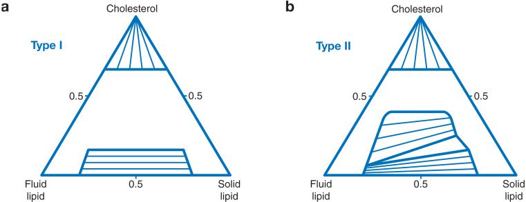
Bilayer mixtures of lipids are used by many researchers as chemically simple models for biological membranes. In particular, observations on three-component bilayer mixtures containing cholesterol show rich phase behavior, including several regions of two-phase coexistence and one region of three-phase coexistence. Yet, the relationship between these simple model mixtures and biological membranes, which contain hundreds of different proteins and lipids, is not clear. Many of the model mixtures have been chosen for study because they exhibit readily observed phase separations, not because they are good mimics of cell membrane components. If the many components of cell membranes could be grouped in some way, then understanding the phase behaviors of biological membranes might be enhanced. Furthermore, if the underlying interaction energies between lipids and proteins can be determined, then it might be possible to model the distributions of lipids and proteins in a bilayer membrane, even in complex mixtures.
Figures
References
-
- Andersen OS, Koeppe RE., II Bilayer thickness and membrane protein function. Annu. Rev. Biophys. Biomol. Struct. 2006;36 In press. - PubMed
-
- Anderson RGW, Jacobson K. A role for lipid shells in targeting proteins to caveolae, rafts, and other lipid domains. Science. 2002;296:1821–25. - PubMed
-
- Angelova MI, Soleau S, Meleard P, Faucon JF, Bothorel P. Preparation of giant vesicles by external AC electric fields. Kinetics and applications. Prog. Colloid Polym. Sci. 1992;89:127–31.
Publication types
MeSH terms
Substances
Grants and funding
LinkOut - more resources
Full Text Sources