Review
. 2007:25:619-47.
doi: 10.1146/annurev.immunol.25.022106.141618.

Structural basis of integrin regulation and signaling

Affiliations
Review

Structural basis of integrin regulation and signaling

Bing-Hao Luo et al. Annu Rev Immunol. 2007.

Abstract

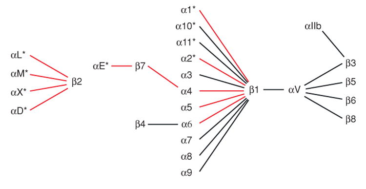
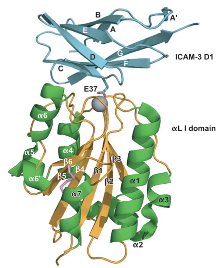
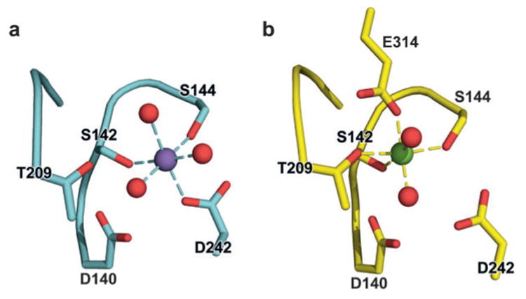
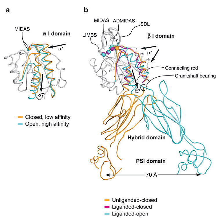
Integrins are cell adhesion molecules that mediate cell-cell, cell-extracellular matrix, and cell-pathogen interactions. They play critical roles for the immune system in leukocyte trafficking and migration, immunological synapse formation, costimulation, and phagocytosis. Integrin adhesiveness can be dynamically regulated through a process termed inside-out signaling. In addition, ligand binding transduces signals from the extracellular domain to the cytoplasm in the classical outside-in direction. Recent structural, biochemical, and biophysical studies have greatly advanced our understanding of the mechanisms of integrin bidirectional signaling across the plasma membrane. Large-scale reorientations of the ectodomain of up to 200 A couple to conformational change in ligand-binding sites and are linked to changes in alpha and beta subunit transmembrane domain association. In this review, we focus on integrin structure as it relates to affinity modulation, ligand binding, outside-in signaling, and cell surface distribution dynamics.


Figures

Figure 1
Figure 1
The 24 integrin heterodimers. The α subunits with α I domains are asterisked. Integrin heterodimers on immune cells are shown with red lines.
Figure 2
Figure 2
A mutant, high-affinity αL I domain (gold β-sheet and coil and green α-helices) in complex with domain 1 of ICAM-3 (cyan). The Mg2+ is shown as a gray sphere. The side chain of the key integrin-binding residue, Glu37 of ICAM-3, is shown. The mutationally introduced K287C/K294C disulfide bond that stabilizes the open conformation is shown in pink. ICAM-3 domain 2 is omitted for clarity. [From Protein Data Bank (PDB) ID code 1T0P (7).]
Figure 3
Figure 3
Structural rearrangement of the αM I domain MIDAS. (a) Structure of the closed αM I domain MIDAS. (b) Structure of the open α I domain MIDAS. Glu-314 from a neighboring αM I domain coordinates with the MIDAS magnesium. Purple and green spheres are Mn2+ and Mg2+ ions, respectively, and red spheres are coordinating water-molecule oxygens. [From PDB ID codes 1JLM and 1IDO (4, 8).]
Figure 4
Figure 4
Conformational change and transmission of allostery by α and β I domains. (a) The α I domain. Nonmoving segments of the backbone are shown as a gray worm. The moving segments of the backbone and the MIDAS metal ions are closed (gold ) and open (cyan). The direction of movement is indicated with arrows. [From PDB ID codes 1JLM and 1IDO (4, 8).] (b) The β I domain and its linkage to the hybrid and plexin/semaphorin/integrin (PSI) domain. Nonmoving segments of the β I backbone are shown as a gray worm. Moving segments and metal ions are color coded as shown. Directions of α1- and α7-helix movements are shown with arrows. [PDB ID codes are 1U8C, 1L5G, and 1TXV (32, 36, 40).]
Figure 5
Figure 5
ICAM structure and integrin binding. (a) Schematic of ICAM-1, -2, -3, and -5. The domains are color coded, and integrin-binding sites are shown. (b) Structural model of ICAM-1 oligomers bound to αL I domain. The model was constructed from the structure of ICAM-1 D1-D2 in complex with αL I domain (PDB ID code 1MQ8) and from the structure of ICAM-1 D3-D5 (PDB ID code 1P53) (6, 24). ICAM-1 is cyan, with the first carbohydrate residue at each site in yellow; the αL I domain is purple.
Figure 6
Figure 6
Integrin architecture. (a) Organization of domains within the primary structures. Some α subunits contain an I domain inserted in the position denoted by the dashed lines. Cysteines and disulfides are shown as lines below the stick figures. (b) Schematic of the course of the α and β subunit polypeptide chains through domains from the N to C termini. (c-d ) Rearrangement of domains during activation of integrins that lack (c) or contain (d ) an α I domain. The β subunit lower legs are flexible and are therefore shown in what may be the predominant (solid representation) and less predominant (dashed lines) orientations.
Figure 7
Figure 7
Crystal structures of integrins αVβ3 and αIIbβ3. (a) The structure of αVβ3 in the bent conformation. The αV and β3 subunits are colored in green and red, respectively. (b) Superposition of liganded-open αIIbβ3 and unliganded-closed αVβ3 headpieces. The α and β subunits are colored in green and yellow in αVβ3 and in purple and light blue in αIIbβ3. Calcium and magnesium ions in αIIbβ3 only are gold and gray spheres, respectively. (c) The drug tirofiban is shown bound to the αIIbβ3 head, and mapping is shown of fibrinogen binding-sensitive mutations. The clinically approved antagonist tirofiban is shown with yellow carbons, blue nitrogens, and red oxygens. The cap subdomain of the β-propeller is in green. Ca2+ and Mg2+ ions are large gold and gray spheres, respectively. Cβ atoms of fibrinogen binding-sensitive residues are shown as small spheres in the same color as the domains in which they are present. Disulfide bonds are yellow cylinders. (d ) The binding of eptifibatide to αIIbβ3 interface is depicted. The fragment of eptifibatide that mimics RGD is shown as a stick model with carbon, nitrogen, and oxygen colored yellow, blue, and red, respectively. The binding pocket is shown with αIIb and β3 in purple and light blue, respectively. Hydrogen bonds are shown as gray dashed lines. Ca2+ and Mg2+ are gold and gray spheres, respectively. The coordinations to the metal ions are shown as green dashed lines. [Structure PDB ID codes are, for αVβ3, 1U8C (40); for αIIbβ3 bound to eptifibatide, 1TY6; and for αIIbβ3 bound to tirofiban, 1TY5 (36).]
Figure 8
Figure 8
Communication between α I and β I domains. (a) It has been proposed that αL-Glu-310 acts as an intrinsic ligand that binds to the β2 I domain MIDAS and, thus, axially displaces the αL I domain α7-helix in the C-terminal direction, reshapes the β6-α7 loop, and activates the αL I domain MIDAS. (b) Individual mutation of αL-Glu-310 or β2-Ala-210 to cysteine abolishes I domain activation, whereas the double mutation of αL-E310C with β2-A210C forms a disulfide bond that constitutively activates ligand binding (104).
Figure 9
Figure 9
EM negative-stain class averages of integrins αVβ3 and αXβ2 in bent and extended conformations (33, 43). The EM images of the extended conformations only are colored according to the scheme shown in d. (a) αVβ3 in bent ( panel 1), extended with closed headpiece ( panel 2), and extended with open headpiece ( panel 3) conformations. (b) αXβ2 in bent ( panel 1), extended with closed headpiece ( panel 2), and extended with open headpiece ( panel 3) conformations. (c) αXβ2 in complex with CBR LFA-1/2 Fab illustrates flexibility of the β leg: panel 1, closed headpiece with parallel legs; panel 2, closed headpiece with crossed legs; panels 3 and 4, open headpiece. Panels 1–3 are with clasped αXβ2, and panel 4 is with unclasped αXβ2. In a-c, a schematic in the same orientation as the right-most panel is shown to the right; the dashed lower β legs symbolize flexibility and averaging-out.

Similar articles

Cited by

References

    1. Pribila JT, Quale AC, Mueller KL, Shimizu Y. Integrins and T cell-mediated immunity. Annu Rev Immunol. 2004;22:157–80. - PubMed
    1. Kinashi T. Intracellular signaling controlling integrin activation in lymphocytes. Nat Rev Immunol. 2005;5:546–59. - PubMed
    1. Anderson DC, Springer TA. Leukocyte adhesion deficiency: an inherited defect in the Mac-1, LFA-1, and p150,95 glycoproteins. Annu Rev Med. 1987;38:175–94. - PubMed
    1. Lee JO, Rieu P, Arnaout MA, Liddington R. Crystal structure of the A domain from the α subunit of integrin CR3 (CD11b/CD18) Cell. 1995;80:631–38. - PubMed
    1. Emsley J, Knight CG, Farndale RW, Barnes MJ, Liddington RC. Structural basis of collagen recognition by integrin α2β1. Cell. 2000;101:47–56. - PubMed

Publication types

MeSH terms

LinkOut - more resources