

Expression patterns of CEACAM5 and CEACAM6 in primary and metastatic cancers
- PMID: 17201906
- PMCID: PMC1769503
- DOI: 10.1186/1471-2407-7-2
Expression patterns of CEACAM5 and CEACAM6 in primary and metastatic cancers
Abstract
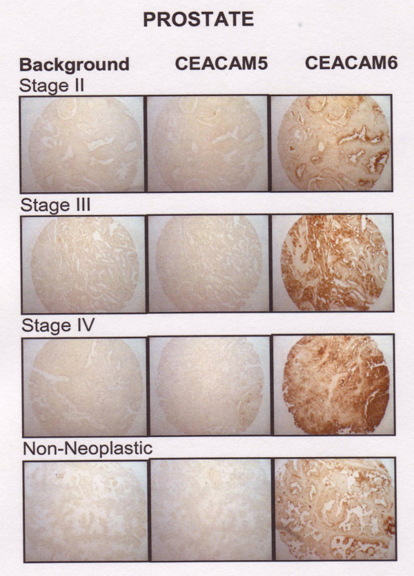
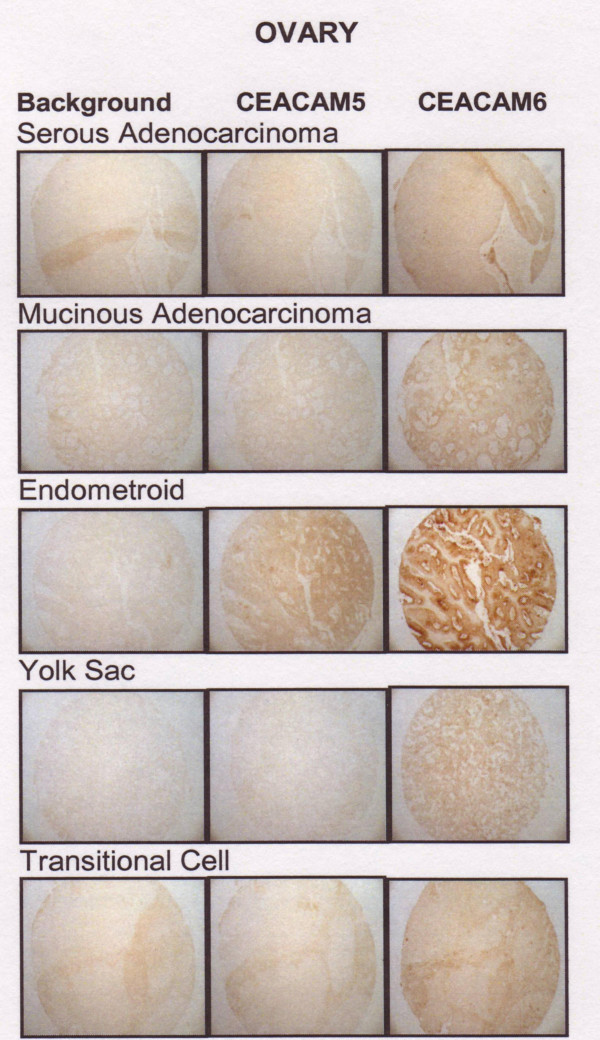
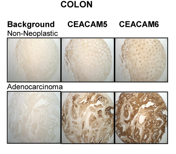
Background: Many breast, pancreatic, colonic and non-small-cell lung carcinoma lines express CEACAM6 (NCA-90) and CEACAM5 (carcinoembryonic antigen, CEA), and antibodies to both can affect tumor cell growth in vitro and in vivo. Here, we compare both antigens as a function of histological phenotype in breast, pancreatic, lung, ovarian, and prostatic cancers, including patient-matched normal, primary tumor, and metastatic breast and colonic cancer specimens.
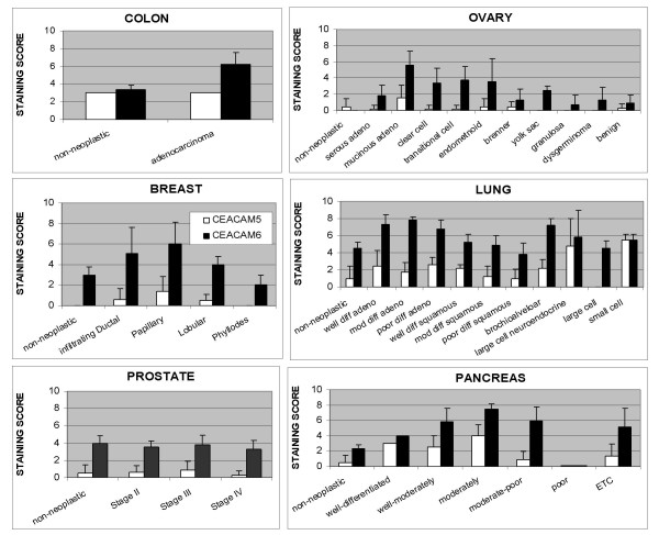
Methods: Antigen expression was determined by immunohistochemistry (IHC) using tissue microarrays with MN-15 and MN-3 antibodies targeting the A1B1- and N-domains of CEACAM6, respectively, and the MN-14 antibody targeting the A3B3 domain of CEACAM5. IHC was performed using avidin-biotin-diaminobenzide staining. The average score +/- SD (0 = negative/8 = highest) for each histotype was recorded.
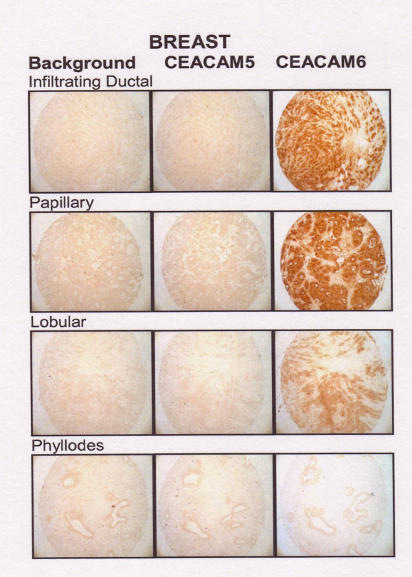
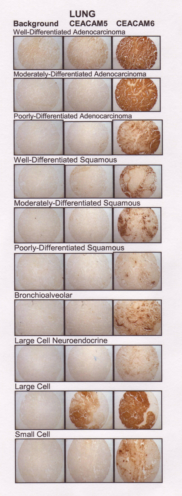
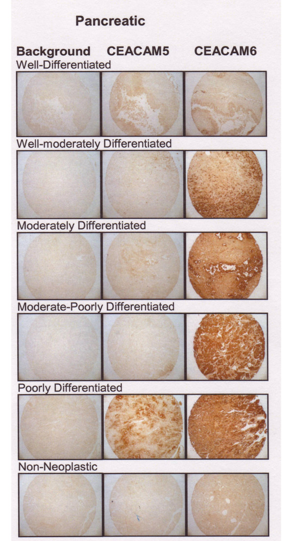
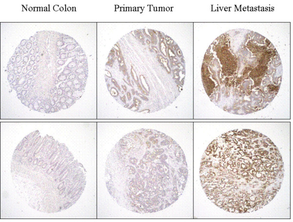
Results: For all tumors, the amount of CEACAM6 expressed was greater than that of CEACAM5, and reflected tumor histotype. In breast tumors, CEACAM6 was highest in papillary > infiltrating ductal > lobular > phyllodes; in pancreatic tumors, moderately-differentiated > well-differentiated > poorly-differentiated tumors; mucinous ovarian adenocarcinomas had almost 3-fold more CEACAM6 than serous ovarian adenocarcinomas; lung adenocarcinomas > squamous tumors; and liver metastases of colonic carcinoma > primary tumors = lymph nodes metastases > normal intestine. However, CEACAM6 expression was similar in prostate cancer and normal tissues. The amount of CEACAM6 in metastatic colon tumors found in liver was higher than in many primary colon tumors. In contrast, CEACAM6 immunostaining of lymph node metastases from breast, colon, or lung tumors was similar to the primary tumor.
Conclusion: CEACAM6 expression is elevated in many solid tumors, but variable as a function of histotype. Based on previous work demonstrating a role for CEACAM6 in tumor cell migration, invasion and adhesion, and formation of distant metastases (Blumenthal et al., Cancer Res 65: 8809-8817, 2005), it may be a promising target for antibody-based therapy.
Figures
Similar articles
-
Inhibition of adhesion, invasion, and metastasis by antibodies targeting CEACAM6 (NCA-90) and CEACAM5 (Carcinoembryonic Antigen).Cancer Res. 2005 Oct 1;65(19):8809-17. doi: 10.1158/0008-5472.CAN-05-0420. Cancer Res. 2005. PMID: 16204051
-
Overexpression and clinical significance of carcinoembryonic antigen-related cell adhesion molecule 6 in colorectal cancer.Clin Chim Acta. 2013 Jan 16;415:12-9. doi: 10.1016/j.cca.2012.09.003. Epub 2012 Sep 10. Clin Chim Acta. 2013. PMID: 22975528
-
Carcinoembryonic antigen-related cell adhesion molecule 1 is expressed and as a function histotype in ovarian tumors.Ann Diagn Pathol. 2016 Feb;20:7-12. doi: 10.1016/j.anndiagpath.2015.10.012. Epub 2015 Oct 27. Ann Diagn Pathol. 2016. PMID: 26653024
-
Carcinoembryonic antigen-related cell adhesion molecules (CEACAMs) in cancer progression and metastasis.Cancer Metastasis Rev. 2013 Dec;32(3-4):643-71. doi: 10.1007/s10555-013-9444-6. Cancer Metastasis Rev. 2013. PMID: 23903773 Review.
-
Towards understanding the mechanisms of actions of carcinoembryonic antigen-related cell adhesion molecule 6 in cancer progression.Cancer Sci. 2018 Jan;109(1):33-42. doi: 10.1111/cas.13437. Epub 2018 Jan 2. Cancer Sci. 2018. PMID: 29110374 Free PMC article. Review.
Cited by
-
Dual-inhibitory domain iCARs improve the efficiency of the AND-NOT gate CAR T strategy.Proc Natl Acad Sci U S A. 2023 Nov 21;120(47):e2312374120. doi: 10.1073/pnas.2312374120. Epub 2023 Nov 14. Proc Natl Acad Sci U S A. 2023. PMID: 37963244 Free PMC article.
-
Biomarkers in bile-complementing advanced endoscopic imaging in the diagnosis of indeterminate biliary strictures.World J Gastrointest Endosc. 2015 Apr 16;7(4):308-17. doi: 10.4253/wjge.v7.i4.308. World J Gastrointest Endosc. 2015. PMID: 25901209 Free PMC article. Review.
-
Proteomic alterations in early stage cervical cancer.Oncotarget. 2018 Apr 6;9(26):18128-18147. doi: 10.18632/oncotarget.24773. eCollection 2018 Apr 6. Oncotarget. 2018. PMID: 29719595 Free PMC article.
-
Carcinoembryonic antigen-related cell adhesion molecules (CEACAM) 1, 5 and 6 as biomarkers in pancreatic cancer.PLoS One. 2014 Nov 19;9(11):e113023. doi: 10.1371/journal.pone.0113023. eCollection 2014. PLoS One. 2014. PMID: 25409014 Free PMC article.
-
Design and activity of a murine and humanized anti-CEACAM6 single-chain variable fragment in the treatment of pancreatic cancer.Cancer Res. 2009 Mar 1;69(5):1933-40. doi: 10.1158/0008-5472.CAN-08-2707. Epub 2009 Feb 24. Cancer Res. 2009. PMID: 19244123 Free PMC article.
References
-
- Beauchemin N, Draber P, Dveksler G, Gold P, Gray-Owen S, Grunert F, Hammerstrom S, Holmes KV, Karlsson A, Kuroki M, Lin SH, Lucka L, Najjar SM, Neumaier M, Obrink B, Shively JE, Skubitz KM, Stanners CP, Thomas P, Thompason JA, Virji M, von Kleist S, Wagener C, Watt S, Zimmermann W. Redefined nomenclature for members of the carcinoembryonic antigen family. Exp Cell Res. 1999;252:243–249. doi: 10.1006/excr.1999.4610. - DOI - PubMed
-
- Hammarstrom S, Olsen A, Teglund S, Baranov V. The nature and expression of the human CEA family. In: Stanners CO, editor. Cell Adhesion and Communications Mediated by the CEA Family Basic and Clinical Perspectives. Vol. 5. Amsterdam, Harwood Academic Publishers; 1998. pp. 1–30.
-
- Goldenberg DM, Sharkey RM, Primus FJ. Carcinoembryonic antigen in histopathology: immunoperoxidase staining of conventional tissue sections. J Natl Cancer Inst. 1976;57:11–22. - PubMed
-
- Shively JE, Beatty JD. CEA-related antigens: molecular biology and clinical significance. Crit Rev Oncol Hematol. 1985;2:355–399. - PubMed
Publication types
MeSH terms
Substances
Grants and funding
LinkOut - more resources
Full Text Sources
Other Literature Sources
