

Expression of Cre recombinase in dopaminoceptive neurons
- PMID: 17201924
- PMCID: PMC1770923
- DOI: 10.1186/1471-2202-8-4
Expression of Cre recombinase in dopaminoceptive neurons
Abstract
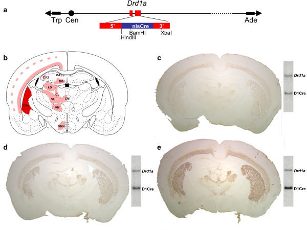
Background: Dopamine-activated signaling regulates locomotor and emotional responses and alterations in dopamine-signaling are responsible of several psychomotor disorders. In order to identify specific functions of these pathways, the Cre/loxP system has been used. Here, we describe the generation and the characterization of a transgenic mouse line expressing the Cre recombinase in dopaminoceptive neurons. To this purpose, we used as expression vector a 140 kb yeast artificial chromosome (YAC) containing the dopamine D1 receptor gene (Drd1a).
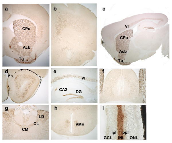
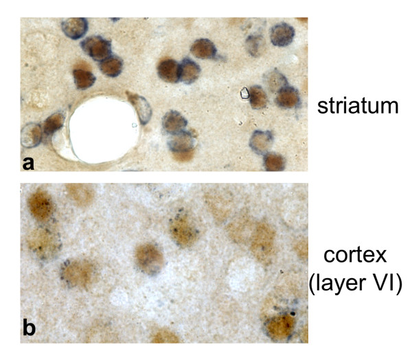
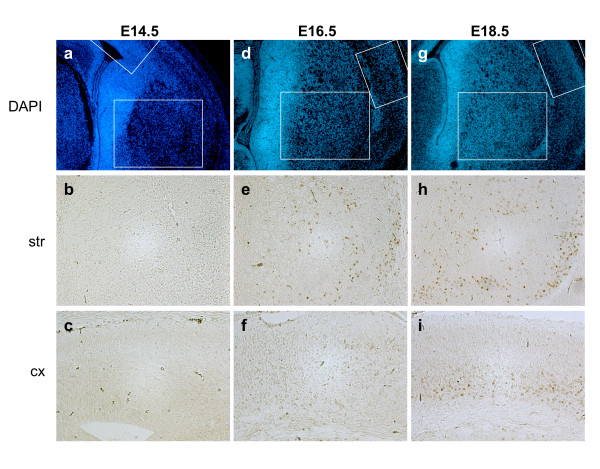
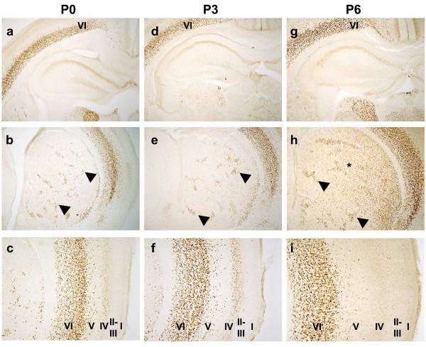
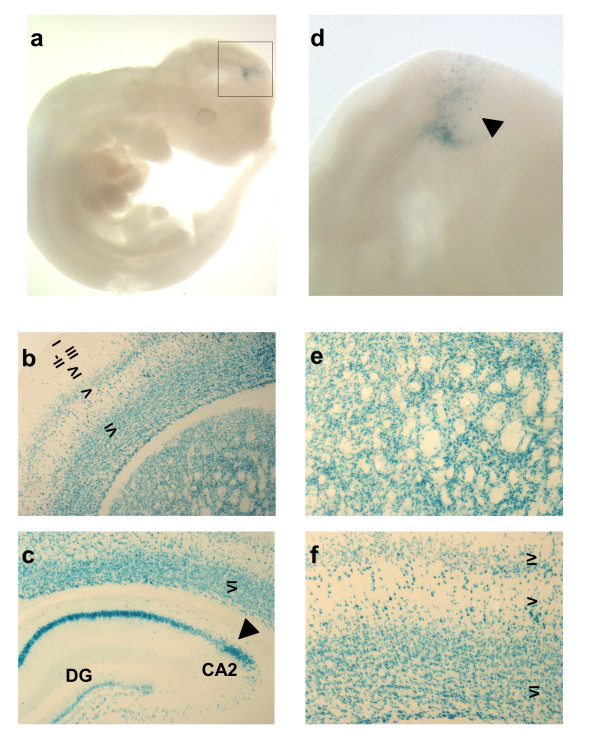
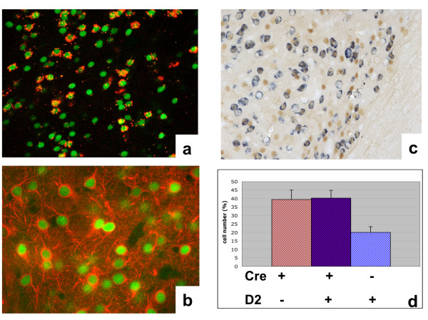
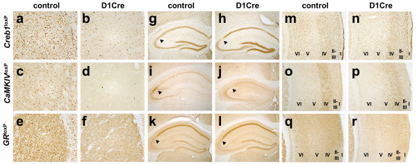
Results: In the chosen line, D1Cre, the spatio-temporal pattern of Cre expression closely recapitulated that of the endogenous Drd1a gene, as assessed by immunohistological approaches in embryonic and adult stages. Efficiency of recombination was confirmed by crossing D1Cre with three different loxP lines (Creb1loxP, CaMKIVloxP and GRloxP) and with the R26R reporter. In the three loxP lines studied, recombination was restricted to the area of Cre expression.
Conclusion: In view of the patterns of recombination restricted to the major dopaminoceptive regions as seen in the context of the CREB, CaMKIV and GR mutations, the D1Cre line will be a useful tool to dissect the contributions of specific genes to biological processes involving dopamine signaling.
Figures
Similar articles
-
Inducible gene inactivation in neurons of the adult mouse forebrain.BMC Neurosci. 2007 Aug 2;8:63. doi: 10.1186/1471-2202-8-63. BMC Neurosci. 2007. PMID: 17683525 Free PMC article.
-
Transgenic mice engineered to target Cre/loxP-mediated DNA recombination into catecholaminergic neurons.Genesis. 2003 Aug;36(4):196-202. doi: 10.1002/gene.10217. Genesis. 2003. PMID: 12929090
-
Temporal regulation of Cre-recombinase activity in Scl-positive neurons of the central nervous system.Genesis. 2007 Mar;45(3):145-51. doi: 10.1002/dvg.20274. Genesis. 2007. PMID: 17330263
-
Conditional gene targeting in the mouse nervous system: Insights into brain function and diseases.Pharmacol Ther. 2007 Mar;113(3):619-34. doi: 10.1016/j.pharmthera.2006.12.003. Epub 2007 Jan 10. Pharmacol Ther. 2007. PMID: 17289150 Review.
-
Production of a reporter transgenic pig for monitoring Cre recombinase activity.Biochem Biophys Res Commun. 2009 May 1;382(2):232-5. doi: 10.1016/j.bbrc.2009.02.146. Epub 2009 Mar 5. Biochem Biophys Res Commun. 2009. PMID: 19268654 Review.
Cited by
-
Dopamine inhibits mitochondrial motility in hippocampal neurons.PLoS One. 2008 Jul 30;3(7):e2804. doi: 10.1371/journal.pone.0002804. PLoS One. 2008. PMID: 18665222 Free PMC article.
-
Dicer loss in striatal neurons produces behavioral and neuroanatomical phenotypes in the absence of neurodegeneration.Proc Natl Acad Sci U S A. 2008 Apr 8;105(14):5614-9. doi: 10.1073/pnas.0801689105. Epub 2008 Apr 2. Proc Natl Acad Sci U S A. 2008. PMID: 18385371 Free PMC article.
-
The striatal balancing act in drug addiction: distinct roles of direct and indirect pathway medium spiny neurons.Front Neuroanat. 2011 Jul 18;5:41. doi: 10.3389/fnana.2011.00041. eCollection 2011. Front Neuroanat. 2011. PMID: 21811439 Free PMC article.
-
Stress and addiction: glucocorticoid receptor in dopaminoceptive neurons facilitates cocaine seeking.Nat Neurosci. 2009 Mar;12(3):247-9. doi: 10.1038/nn.2282. Epub 2009 Feb 22. Nat Neurosci. 2009. PMID: 19234455
-
Development of a BAC vector for integration-independent and tight regulation of transgenes in rodents via the Tet system.Transgenic Res. 2011 Jun;20(3):709-20. doi: 10.1007/s11248-010-9427-0. Epub 2010 Jul 18. Transgenic Res. 2011. PMID: 20640885
References
-
- Houk JC, Davis JL, Beiser DG. Models of Information Processing in the Basal Ganglia. , MIT Press; 1994. p. 390.
-
- Nieoullon A, Coquerel A. Dopamine: a key regulator to adapt action, emotion, motivation and cognition. Curr Opin Neurol. 2003;16 Suppl 2:S3–9. - PubMed
Publication types
MeSH terms
Substances
LinkOut - more resources
Full Text Sources
Other Literature Sources
Molecular Biology Databases
