. 2007 Mar;81(6):2592-604.
doi: 10.1128/JVI.01643-06. Epub 2007 Jan 3.

High-frequency epigenetic repression and silencing of retroviruses can be antagonized by histone deacetylase inhibitors and transcriptional activators, but uniform reactivation in cell clones is restricted by additional mechanisms

Affiliations

High-frequency epigenetic repression and silencing of retroviruses can be antagonized by histone deacetylase inhibitors and transcriptional activators, but uniform reactivation in cell clones is restricted by additional mechanisms

Richard A Katz et al. J Virol. 2007 Mar.

Abstract

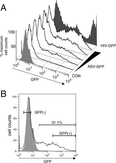
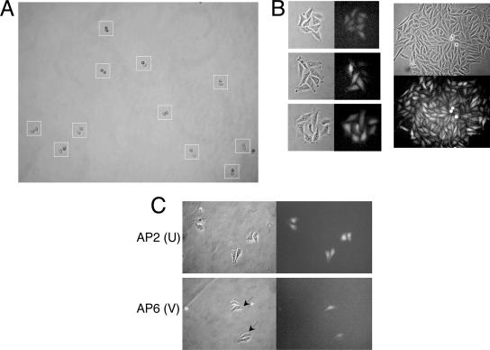
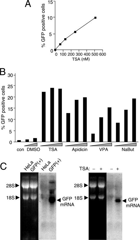
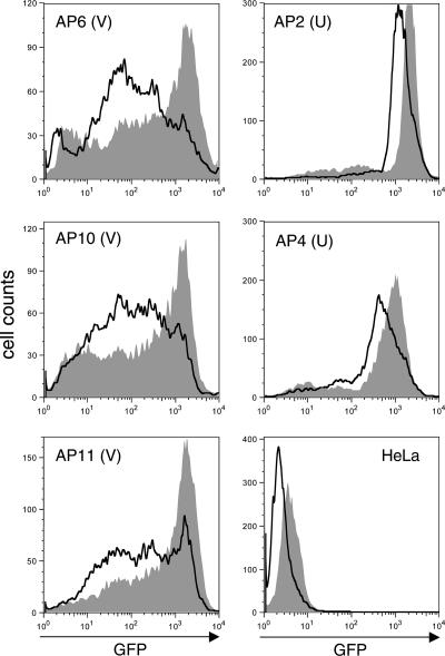
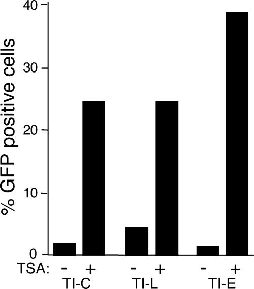
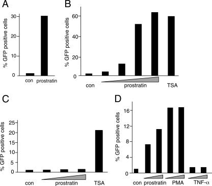
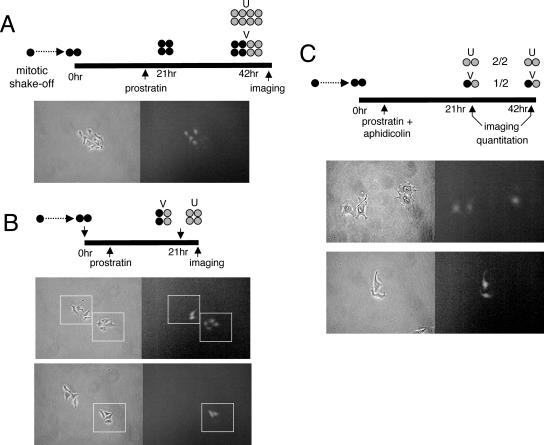
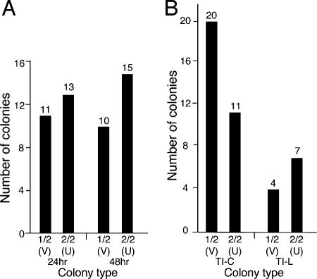
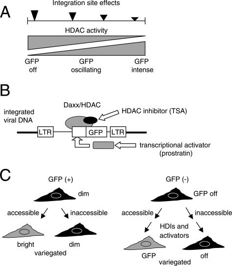
Integrated retroviral DNA is subject to epigenetic gene silencing, but the viral and host cell properties that influence initiation, maintenance, and reactivation are not fully understood. Here we describe rapid and high-frequency epigenetic repression and silencing of integrated avian sarcoma virus (ASV)-based vector DNAs in human HeLa cells. Initial studies utilized a vector carrying the strong human cytomegalovirus (hCMV) immediate-early (IE) promoter to drive expression of a green fluorescent protein (GFP) reporter gene, and cells were sorted into two populations based on GFP expression [GFP(+) and GFP(-)]. Two potent epigenetic effects were observed: (i) a very broad distribution of GFP intensities among cells in the GFP(+) population as well as individual GFP(+) clones and (ii) high-frequency GFP reporter gene silencing in GFP(-) cells. We previously showed that histone deacetylases (HDACs) can associate with ASV DNA soon after infection and may act to repress viral transcription at the level of chromatin. Consistent with this finding, we report here that treatment with the histone deacetylase inhibitor trichostatin A (TSA) induces GFP activation in GFP(-) cells and can also increase GFP expression in GFP(+) cells. In the case of the GFP(-) populations, we found that after removal of TSA, GFP silencing was reestablished in a subset of cells. We used that finding to enrich for stable GFP(-) cell populations in which viral GFP reporter expression could be reactivated by TSA; furthermore, we found that the ability to isolate such populations was independent of the promoter driving the GFP gene. In such enriched cultures, hCMV IE-driven, but not the viral long terminal repeat-driven, silent GFP reporter expression could be reactivated by the transcriptional activator prostratin. Microscopy-based studies using synchronized cells revealed variegated reactivation in cell clones, indicating that secondary epigenetic effects can restrict reactivation from silencing. Furthermore we found that entry into S phase was not required for reactivation. We conclude that HDACs can act rapidly to initiate and maintain promoter-independent retroviral epigenetic repression and silencing but that reactivation can be restricted by additional mechanisms.


Figures

FIG. 1.
FIG. 1.
GFP intensity profiles of infected HeLa cells as analyzed by FACS. (A) Profiles of GFP expression were measured at 48 h postinfection. Shown are GFP profiles for uninfected HeLa cells (CON) (light shading) and for cells after infection with increasing amounts of the ASV-GFP vector. A parallel culture was infected with an HIV-1-based vector (HIV-GFP) (dark shading). Data were analyzed using FlowJo software. y-axis scaling was used to more readily compare the intensity profiles. (B) Gating strategy to isolate GFP(−) and GFP(+) cells. A representative profile from panel A is shown with the approximate gates used for isolation of cell populations by preparative cell sorting. In this example, the GFP(+) cells represented ca. 30% of the culture. A subfraction of brighter GFP(+) cells were selected for isolation to ensure that GFP patterns were bright enough to observe microscopically.
FIG. 2.
FIG. 2.
Variegation of GFP expression during outgrowth of GFP(+) microclones. (A) A mitotic shakeoff strategy was used to promote highly synchronized outgrowth of microclones from single cells. Shown is a single field, imaged ca. 2 h after plating mitotic cells. Typically, synchronization occurred with 70% to 90% efficiency. Boxed colonies have completed cytokinesis synchronously and entered G1 as indicated by the cell doublets. (B) GFP patterns were monitored during colony outgrowth. Left panels show eight-cell colonies, with the upper panel illustrating a colony displaying uniform GFP expression and the middle and bottom panels highlighting variegated colonies. Right panels show a representative variegated pattern in a large colony. Phase-contrast and fluorescent images are shown. (C) Analysis of GFP variegation during outgrowth of synchronous microclones derived from GFP(+) cell clones. One uniform (U) (AP2) and one variegating (V) (AP6) clone were examined. Representative images are shown. Arrows indicate cells displaying higher-intensity GFP expression.
FIG. 3.
FIG. 3.
Reactivation of GFP reporter genes after treatment with HDIs. (A) Cells were sorted at 8 days postinfection, and the GFP(−) cells were passaged for several months. GFP(−) cells were treated with the indicated concentrations of TSA, and GFP expression was quantitated by FACS at 24 h posttreatment. (B) TI-C cultures that were passaged for several months were challenged with the indicated HDIs, and GFP expression was quantitated by FACS at 24 h posttreatment. con, not treated. Concentrations: dimethyl sulfoxide (DMSO), 0.05% and 0.2%; TSA, 0.5, 1, and 2 μM; apidicin, 0.5, 1, and 2 μg/ml; valproic acid (VPA), 2, 4, and 8 mM; sodium butyrate (NaBut), 2, 4, and 8 mM. (C) Northern blot analysis of GFP mRNA was carried out using standard methods. GFP mRNA was characterized in a pool of GFP(+) HeLa cells infected with the ASV construct in which the GFP gene is under control of the hCMV IE promoter (left panel). RNA loading was monitored by staining of 18S and 28S RNAs, and these species also served as sizing standards (5,025 and 1,868 nucleotides, respectively). A GFP transcript of ca. 1,700 nucleotides was identified, and this size is consistent with initiation within the internal hCMV viral promoter and 3′ processing at the 3′ LTR. This transcript was not detected in TI-C cells but was induced to significant levels after treatment with TSA (1 μM) (right panel).
FIG. 4.
FIG. 4.
FACS analysis of GFP(+) cell clones after treatment with TSA. Eleven GFP(+) clones were categorized as variegated (V) or uniform (U) by microscopy. Analyses of several representative clones (V clones, AP6, AP10, and AP11; U clones, AP2 and AP4) are presented. Cultures were treated with 0.5 μM TSA for 24 h and analyzed by FACS. Nonspecific effects were monitored by treatment of uninfected HeLa cells (HeLa). Data were processed with FlowJo software. Untreated, no fill; TSA treated, filled.
FIG. 5.
FIG. 5.
Characterization of cell populations enriched for TSA-inducible hCMV IE-, ASV LTR-, and EF1 alpha-driven GFP expression. The indicated cell populations were treated with TSA (1 μM) for 24 h, and GFP expression was quantitated by FACS analysis. TI-C, hCMV promoter; TI-L, ASV LTR promoter; TI-E, EF-1α promoter.
FIG. 6.
FIG. 6.
Phorbol esters can reactivate silent GFP reporter gene expression in TSA-inducible TI-C but not TI-L populations. GFP expression in treated and untreated (con) cells was quantitated by FACS analysis at 24 h posttreatment. (A) A Jurkat cell clone (10.6) harboring latent HIV encoding a GFP reporter (30, 62) was treated with prostratin (2 μM). (B) TI-C cells were treated with prostratin (0.1, 0.3, 1.0, and 1.5 μM) or TSA (2 μM). (C) TI-L cells were treated with prostratin (0.3, 1.0, and 1.5 μM) or TSA (2 μM). (D) TI-C cells were treated with prostratin (2.0, 5.0 μM), PMA (100 and 200 nM), and TNF-α (10, 20 ng/ml).
FIG. 7.
FIG. 7.
Analysis of GFP reactivation patterns during clonal outgrowth of TI-C microclones reveals a variegated response. TI-C cell populations were synchronized by mitotic shakeoff and plated under dilute conditions. Colonies were treated at various times postplating with prostratin, and fluorescence (right) and phase-contrast (left) images were acquired as indicated in the diagrams: black fill, nonexpressing cells; gray fill, GFP-expressing cells. (A) Treatment at the two-cell stage resulted in variegated (V) as well as uniform (U) GFP expression at the eight-cell stage. Shown is a representative colony displaying variegated expression. (B) The two-cell colonies were treated with prostratin at 2 h postplating, and colonies were imaged at the four-cell stage (boxed). Variegated and uniform expression patterns were observed. The top panel shows neighboring V- and U-type colonies, and the bottom panel shows a V-type colony. Cell-to-cell intensity of GFP expression is constant within each clone (boxed). (C) Treatment was similar to that for panel B, except that aphidicolin was included to prevent cells from progressing into S phase. Colonies were arrested at the two-cell stage and could be quantitated according to V (1/2) or U (2/2) patterns (see Fig. 8). Representative images are shown.
FIG. 8.
FIG. 8.
Quantitation of variegated (V) and uniform (U) GFP expression in two-cell colonies after aphidicolin arrest and reactivation with prostratin (1 μM) or TSA (0.5 μM). The experimental design is shown in Fig. 7C, and representative results are shown. (A) TI-C 2-cell colonies were quantitated for V or U patterns at 24 and 48 h after plating and prostratin treatment. (B) Comparison of TI-C and TI-L colonies induced with TSA. The sample numbers are lower with TI-L cells, as LTR-driven GFP expression is generally weaker and thus more difficult to detect by microscopy.
FIG. 9.
FIG. 9.
Models for epigenetic silencing and repression. (A) Model for a continuum of HDAC-mediated repression and silencing in GFP(−) and GFP(+) cells. The model is based on the broad GFP expression profiles and the stimulation of GFP expression in both GFP(−) and GFP(+) variegated (V) clones by TSA. Filled arrowheads indicate strong (large arrowhead) or weak (smaller arrowheads) HDAC effects that are modulated by the integration site. (B) Model showing interaction between Daxx/HDAC and integrated retroviral DNA (22). TI cells were selected for the ability of the GFP reporter gene to be reactivated by TSA, an HDI. In such selected cells, phorbol esters can also reactivate the GFP reporter gene, suggesting that the silent loci can be available to transcription factors (rectangle). Coactivator complexes containing HATs may overcome or displace HDACs. (C) The model indicates a common mechanism underlying two phenomena: variegation in some GFP(+) clones and variegated reactivation in GFP(−) TI cells. Expression could be restricted in one daughter cell due to transient inaccessibility of the integration site locus to transcriptional centers (48).

Similar articles

Cited by

References

    1. Barsov, E. V., and S. H. Hughes. 1996. Gene transfer into mammalian cells by a Rous sarcoma virus-based retroviral vector with the host range of the amphotropic murine leukemia virus. J. Virol. 70:3922-3929. - PMC - PubMed
    1. Baur, J. A., J. W. Shay, and W. E. Wright. 2004. Spontaneous reactivation of a silent telomeric transgene in a human cell line. Chromosoma 112:240-246. - PubMed
    1. Berberich, S. L., M. Macias, L. Zhang, L. P. Turek, and C. M. Stoltzfus. 1990. Comparison of Rous sarcoma virus RNA processing in chicken and mouse fibroblasts: evidence for double-spliced RNA in nonpermissive mouse cells. J. Virol. 64:4313-4320. - PMC - PubMed
    1. Bushman, F., M. Lewinski, A. Ciuffi, S. Barr, J. Leipzig, S. Hannenhalli, and C. Hoffmann. 2005. Genome-wide analysis of retroviral DNA integration. Nat. Rev. Microbiol. 3:848-858. - PubMed
    1. Cantrell, S. R., and W. A. Bresnahan. 2006. Human cytomegalovirus (HCMV) UL82 gene product (pp71) relieves hDaxx-mediated repression of HCMV replication. J. Virol. 80:6188-6191. - PMC - PubMed

Publication types