. 2007 Apr;81(7):3109-23.
doi: 10.1128/JVI.02124-06. Epub 2007 Jan 3.

Human cytomegalovirus UL38 protein blocks apoptosis

Affiliations

Human cytomegalovirus UL38 protein blocks apoptosis

Scott Terhune et al. J Virol. 2007 Apr.

Abstract

Apoptosis is an innate cellular defense response to viral infection. The slow-replicating human cytomegalovirus (HCMV) blocks premature death of host cells prior to completion of the infection cycle. In this study, we report that the HCMV UL38 gene encodes a cell death inhibitory protein. A mutant virus lacking the pUL38 coding sequence, ADdlUL38, grew poorly in human fibroblasts, failed to accumulate viral DNA to wild-type levels, and induced excessive death of infected cells. Cells expressing pUL38 were resistant to cell death upon infection and effectively supported the growth of ADdlUL38. Cells infected with the pUL38-deficient virus showed morphological changes characteristic of apoptosis, including cell shrinkage, membrane blebbing, vesicle release, and chromatin condensation and fragmentation. The proteolytic cleavage of two key enzymes involved in apoptosis, namely, caspase 3 and poly(ADP-ribose) polymerase, was activated upon ADdlUL38 infection, and the cleavage was blocked in cells expressing pUL38. The pan-caspase inhibitor Z-VAD-FMK largely restored the growth of ADdlUL38 in normal fibroblasts, indicating that the defective growth of the mutant virus mainly resulted from premature death of host cells. Furthermore, cells expressing pUL38 were resistant to cell death induced by a mutant adenovirus lacking the antiapoptotic E1B-19K protein or by thapsigargin, which disrupts calcium homeostasis in the endoplasmic reticulum. Taken together, these results indicate that the HCMV protein pUL38 suppresses apoptosis, blocking premature death of host cells to facilitate efficient virus replication.


Figures

FIG. 1.
FIG. 1.
Construction of UL38 recombinant BAC-HCMV clones. (A) Viral genomic region carrying UL36-UL38. The first line represents the schematic structure of the viral genomic sequence. The coding sequences of pUL36, pUL37, and pUL38 are indicated by block arrows. The C terminus of each ORF is indicated by the direction of the arrow. Also indicated are the locations of two poly(A) signals immediately downstream of the pUL38 and pUL36 coding sequences and four primers used for PCR amplification to validate sequence alteration introduced in UL38 recombinant BAC-HCMV clones. The 3′ ends of the primers are indicated by the directions of the arrows. The next five lines represent UL36-, UL37-, and UL38-related transcripts. The 3′ end of each transcript is indicated by the direction of the arrow. (B) EcoRI and BamHI restriction digestion analysis. (C) PCR and Southern blot analysis of UL38 recombinant BAC-HCMV clones. Each recombinant BAC-HCMV clone analyzed is described in the text. Dots indicate restriction fragments unique to a recombinant BAC clone due to engineered sequence alterations. For PCR analysis, primer pairs used to amplify specific viral sequences are indicated. For Southern blot analysis, a 32P-labeled probe against the UL38 open reading frame was used to hybridize EcoRI- or BamHI-digested BAC-HCMV DNA. Molecular size markers are indicated.
FIG. 2.
FIG. 2.
The marked growth defect of ADdlUL38 is restored by the expression of pUL38 in trans. Multistep growth analysis was performed with ADwt, ADdlUL38, and ADrevUL38 in normal HF cells (A) and in HF cells transduced with the pUL38-expressing retroviral vector (B). Cells were infected at a multiplicity of 0.01 PFU/cell, culture medium was collected at the indicated times, and yields of cell-free virus were titrated in duplicate by a plaque assay with HF-UL38 cells. Single-step growth analysis of ADdlUL38 in MRC-5 cells was performed to examine the production of extracellular virus (C) and intracellular virus (D). MRC-5 cells were infected with ADdlUL38 or ADwt at an input genome number equivalent to 0.2 PFU of wild-type virus/cell. Infected cell culture medium was collected as extracellular virus samples, and intracellular virus was isolated by freezing and thawing cell pellets. The amount of virus present in each sample was determined by counting the number of IE1-positive cells. The experiment was completed in duplicate.
FIG. 3.
FIG. 3.
Expression and subcellular localization of HCMV pUL38. (A) Fibroblasts were infected with ADdlUL38 or ADwt at an input genome number equivalent to 0.2 PFU of wild-type virus/cell, and cell lysates were analyzed by Western blotting using a pUL38-specific monoclonal antibody at the indicated times. An antibody to alpha-tubulin was used as a loading control. (B) Fibroblasts were infected with ADwt, and immunofluorescence was performed to examine the localization of pUL38 at the indicated times (green). Also shown are the Golgi apparatus, defined by the lectin Helix pomatia agglutinin (red), the nucleus, defined by DAPI (blue), and the merged images.
FIG. 4.
FIG. 4.
ADdlUL38 fails to replicate viral genomes to wild-type levels. (A) Accumulation of viral DNA. Fibroblasts were infected with ADdlUL38 or ADwt at an input genome number equivalent to 0.2 PFU of wild-type virus/cell, total cell-associated DNA was isolated, and viral genomes were quantified using real-time PCR and normalized to β-actin DNA. (B) Accumulation of the UL123 transcript and the protein product IE1. Fibroblasts were infected under the conditions described above. Total RNA was isolated at the indicated times postinfection, and UL123 RNA was quantified by real-time RT-PCR using primers specific to UL123, with SYBR green detection, and normalized to GAPDH RNA. Accumulation of the IE1 protein was determined by Western blot analysis. An antibody to alpha-tubulin was used as a loading control. (C) Accumulation of the UL99 transcript and the protein product pp28. The UL99 transcript was measured by real-time RT-PCR using primers specific to UL99. The pp28 protein was detected by Western blot analysis using an antibody to pp28.
FIG. 5.
FIG. 5.
pUL38 is required to block cell death in HCMV-infected HF cells. (A) For the left panel, normal HF cells were infected with ADwt or ADdlUL38 at an input genome number equivalent to 2 PFU of wild-type virus/cell, and the morphology of infected cells was examined with a phase-contrast microscope at 40 h and 72 h postinfection. For the right panel, quantitative analysis was performed to compare cell death in HF cells infected with ADdlUL38 to that in cells infected with ADwt, as described in Materials and Methods. The ratio of the number of viable cells in ADdlUL38 infection to that in ADwt infection was plotted as a function of hours postinfection (hpi). (B) For the left panel, HF-GFP or HF-UL38 fibroblasts were infected with ADwt or ADdlUL38 at an input genome number equivalent to 1 PFU of wild-type virus/cell, and cell morphology was examined under a phase-contrast microscope at 72 h postinfection. For the right panel, quantitative analysis was performed to compare cell death in various fibroblasts infected with ADdlUL38 to that in cells infected with ADwt, as described in Materials and Methods. The ratio of the number of viable cells in ADdlUL38 infection to that in ADwt infection was plotted for various types of human fibroblasts tested. The results shown are representative of at least two independent experiments.
FIG. 6.
FIG. 6.
Cells infected with the pUL38 deletion mutant demonstrate morphological changes characteristic of apoptosis. (A) Normal HF cells were infected with ADwt-GFP or ADdlUL38-GFP at an input genome number equivalent to 1 PFU of wild-type virus/cell, and the morphology of infected GFP-positive cells was examined under a fluorescence microscope at 72 h and 96 h postinfection. Arrowheads indicate representative cell shrinkage, membrane blebbing, and vesicle release in ADdlUL38-GFP infection. (B) Infected cells were labeled with DAPI at 96 h postinfection. Condensed chromatin in cells infected with ADdlUL38-GFP is indicated by arrowheads. (C) Infected cells were colabeled with DAPI and TUNEL at the indicated times postinfection. The colocalization of GFP, TUNEL, and DAPI staining in the same ADdlUL38-GFP-infected cell population is indicated.
FIG. 7.
FIG. 7.
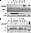
ADdlUL38 induces elevated proteolytic cleavage of caspase 3 and PARP in HF cells. HF-GFP cells were infected with ADwt or ADdlUL38 at an input genome number equivalent to 1 PFU of wild-type virus/cell (A) or HF-GFP and HF-UL38 cells were infected with ADdlUL38 at an input genome number equivalent to 1 PFU of wild-type virus/cell (B), cells were collected at the indicated times postinfection, and cell lysates were examined for cleavage of caspase 3 and PARP by immunoblotting using monoclonal antibodies recognizing only the cleaved forms of the proteins. The infection control was IE1 expression, and the loading control was β-actin expression. Jurkat cell lysates treated with 0.25 mg/ml cytochrome c were used as the positive control for caspase 3 and PARP cleavage.
FIG. 8.
FIG. 8.
Block of cell death by a caspase inhibitor largely restores the growth of ADdlUL38. Normal HF cells were infected with ADwt or ADdlUL38 at an input genome number equivalent to 2 PFU of wild-type virus/cell in the presence or absence of 100 μM Z-VAD-FMK. Culture media of infected cells were collected, and infected cells were supplemented with the appropriate Z-VAD-FMK- or DMSO-containing fresh medium every day for a total of 6 days postinfection. The DMSO-containing medium was used as the solvent control. Yields of cell-free virus were measure in duplicate by a plaque assay with HF-UL38 cells.
FIG. 9.
FIG. 9.
Expression of pUL38 in trans blocks apoptosis induced by the mutant adenovirus dl337. (A) HF-GFP or HF-UL38 cells were infected with dl337. The morphology of infected cells was examined under a phase-contrast microscope (top panel, multiplicity of 3 PFU/cell), and the rate of cell death in dl337-infected cells was quantitated by labeling surviving cells with DAPI and counting DAPI-positive cells (middle panel, 48 h postinfection; bottom panel, multiplicity of 1 PFU/cell). Six independent fields for each sample were counted, and the number of cells in each dl337-infected HF-GFP or HF-UL38 sample was plotted as a percentage of that in a mock-infected sample. (B) HeLa cell lines stably expressing either the empty vector (HeLa-dsRed) or pUL38 (HeLa-UL38) were infected with dl337, the morphology of infected cells was examined under a phase-contrast microscope (top panel, multiplicity of 3 PFU/cell, 40 h postinfection), and the rate of cell death in dl337-infected cells was quantitated as described for panel A (middle panel, 40 h postinfection; bottom panel, multiplicity of 1 PFU/cell). Two independently isolated HeLa-UL38 cell lines (HeLa-UL38-1 and HeLa-UL38-3) were examined.
FIG. 10.
FIG. 10.
Expression of pUL38 in trans blocks ER-mediated apoptosis induced by thapsigargin. pUL38-expressing cells (HF-UL38) or control cells (HF-vector) were treated with 2 μM thapsigargin. Cell morphology was examined under a phase-contrast microscope at 96 h posttreatment (A), and cleavage of caspase 3 was examined by immunoblotting (B). The loading control was β-actin expression. Jurkat cell lysates treated with 0.25 mg/ml cytochrome c were used as the positive control.

Similar articles

Cited by

References

    1. Adair, R., G. W. Liebisch, and A. M. Colberg-Poley. 2003. Complex alternative processing of human cytomegalovirus UL37 pre-mRNA. J. Gen. Virol. 84:3353-3358. - PubMed
    1. Andoniou, C. E., and M. A. Degli-Esposti. 2006. Insights into the mechanisms of CMV-mediated interference with cellular apoptosis. Immunol. Cell Biol. 84:99-106. - PubMed
    1. Arnoult, D., L. M. Bartle, A. Skaletskaya, D. Poncet, N. Zamzami, P. U. Park, J. Sharpe, R. J. Youle, and V. S. Goldmacher. 2004. Cytomegalovirus cell death suppressor vMIA blocks Bax- but not Bak-mediated apoptosis by binding and sequestering Bax at mitochondria. Proc. Natl. Acad. Sci. USA 101:7988-7993. - PMC - PubMed
    1. Benedict, C. A., P. S. Norris, and C. F. Ware. 2002. To kill or be killed: viral evasion of apoptosis. Nat. Immunol. 3:1013-1018. - PubMed
    1. Billstrom Schroeder, M., R. Christensen, and G. S. Worthen. 2002. Human cytomegalovirus protects endothelial cells from apoptosis induced by growth factor withdrawal. J. Clin. Virol. 25(Suppl. 2):S149-S157. - PubMed

Publication types