. 2007 Mar;81(6):2869-79.
doi: 10.1128/JVI.02256-06. Epub 2007 Jan 3.

Cytotoxic T-lymphocyte responses to human papillomavirus type 16 E5 and E7 proteins and HLA-A*0201-restricted T-cell peptides in cervical cancer patients

Affiliations

Cytotoxic T-lymphocyte responses to human papillomavirus type 16 E5 and E7 proteins and HLA-A*0201-restricted T-cell peptides in cervical cancer patients

Dai-Wei Liu et al. J Virol. 2007 Mar.

Abstract

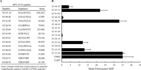
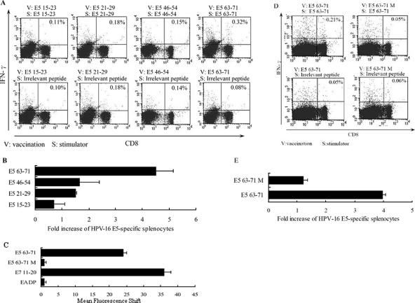
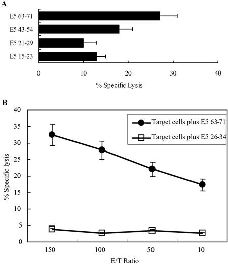
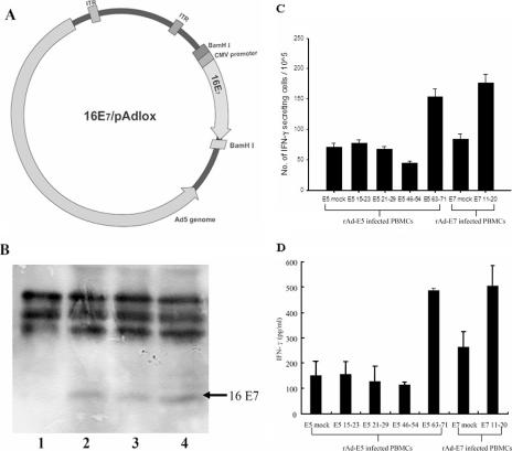
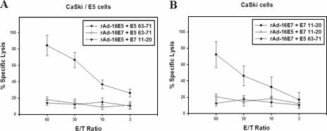
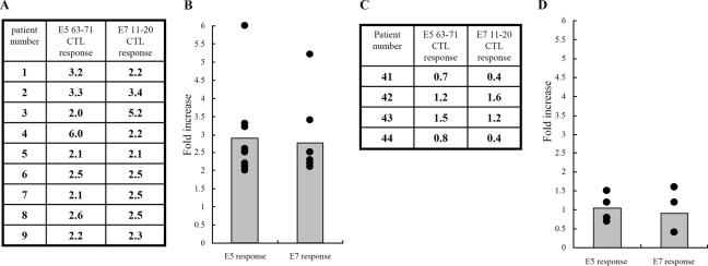
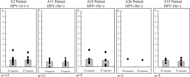
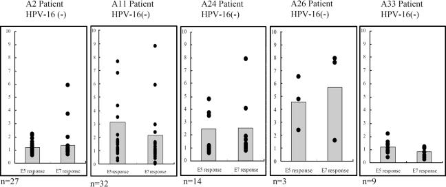
Previously, we found that human papillomavirus type 16 (HPV-16) E5 protein is a tumor rejection antigen and can induce cytotoxic T-lymphocyte (CTL) activity. Therefore, in this study, human leukocyte antigen A*0201 (HLA-A*0201)-restricted human CTL epitopes of HPV-16 E5 protein were identified using a bioinformatics approach, and the abilities of these predicted peptides to induce an immune response in HLA-A*0201 transgenic mice were confirmed by assaying E5-specific CTLs and in vitro-generated CTLs from normal peripheral blood T lymphocytes of HLA-A2-positive human donors. Second, the CTL responses to HLA-A*0201 CTL epitopes (E5 63-71 and E7 11-20) were examined in HPV-16-infected patients with HLA-A2. Third, the effect of HLA-A-type alleles on CTL activities in response to the entire E5 and E7 proteins was examined in cervical cancer patients. E5 and E7 peptides (but not the whole proteins) stimulated E5- and E7-specific CTL recall responses in HPV-16- and HLA-A2-positive cervical cancer patients, and HPV-16 E5 and E7 proteins stimulated naïve T cells in HPV-16-negative cervical cancer patients with HLA-A11 and -A24 haplotypes. In summary, this is the first demonstration that E5 63-71 is an HLA-A*0201-restricted T-cell epitope of HPV-16 E5.


Figures

FIG. 1.
FIG. 1.
Identification of HLA-A*0201-restricted epitopes of HPV-16 E5 protein by T2 cell-binding assays. (A) Computer prediction of HPV-16 E5 peptides. (B) Stabilization of HLA-A*0201 molecules on the surfaces of T2 cells. T2 cells were incubated with peptides overnight at 26°C and then incubated at 37°C for 2 h, as described in Materials and Methods. HLA-A*0201 expression was determined by FACS staining with the monoclonal antibody PA2.1, and the mean fluorescence intensity (MFI) was calculated. The x axis indicates the mean fluorescence shift of T2 cells with tested peptide subtracted from that of T2 cells without peptide. Three experiments were conducted. The data are shown in the form of a histogram. YMDG, the HLA-A*0201 epitope of the tyrosinase-derived peptide YMDGTMSQV, which served as a positive control; E7 11-20, YMLDLQPETT, identified as an HLA-A2-specific CTL epitope of HPV-16 E7 protein, which also served as a positive control; EADP, the HLA-A1 binding peptide EADPTGHSY, which served as a negative control. The MFI values for the positive controls, YMDG and E7 11-20, were 36.8 ± 1.4 and 37.2 ± 7.6, respectively. The MFI value for the negative control, EADP, was 1.93 ± 0.7. The error bars indicate standard deviations.
FIG. 2.
FIG. 2.
Identification of HLA-A*0201-restricted epitopes of HPV-16 E5 protein by vaccination of HLA-A*0201 transgenic mice with peptide plus CpG ODN 1826. Four- to 6-week-old HLA-A*0201 transgenic mice were immunized three times each with T2 cell-binding peptides plus CpG ODN 1826 (12) at 1-week intervals, with five mice in each test group. Five days after the last vaccination, splenocytes were harvested and stimulated with each indicated peptide; then, intracellular-cytokine staining with flow cytometry was performed to determine the number of CD8+ IFN-γ+ double-positive cells. (A) splenocytes from vaccinated mice were stimulated in vitro with the indicated peptide and stained with CD8 and IFN-γ antibodies. The results of one representative assay from three identical independent experiments are shown. The percentage of CD8+ and IFN-γ+ double-positive cells in the gated T-cell populations are shown in the upper corners of the plots. (B) Summary of the three independent experiments. The data represent the means and standard errors of three experiments. The x-axis values were calculated as follows: increase of E5-specific splenocytes = (number of vaccinated splenocytes stimulated with indicated peptide)/(number of vaccinated splenocytes stimulated with irrelevant peptide) × 100%. (C) The T2 cell-binding activities of the wild-type peptide E5 63-71 and the mutant E5 63-71 M. The synthesized wild-type E5 63-71 peptide sequence is YIIFVYIPL, and that of the mutant E5 63-71 is YGIFVYIPG. The measurement of T2 cell binding is described in the legend to Fig. 1B. (D) Splenocytes from vaccinated mice were stimulated in vitro with the E5 63-71 or E5 63-71 M peptide and stained with CD8 and IFN-γ antibodies as described for panel A. (E) Summary of the three independent experiments.
FIG. 3.
FIG. 3.
HPV-16 E5-specific CTL responses in HLA-A*0201 transgenic mice. Four- to 6-week-old HLA-A*0201 transgenic mice were vaccinated three times with each T2 cell-binding peptide plus CpG ODN 1826 at 1-week intervals, with five mice in each test group. Five days after the last vaccination, effector splenocytes were collected and analyzed in in vitro CTL assays as described in Materials and Methods. Target CIR-A2 cells were pulsed with the indicated peptides. (A) CTL activity of each peptide induction. The ratio of effector to target cells from each group of vaccinated mice (five mice) was 100. The data are the average values for five vaccinated mice. The error bars indicate standard deviations. (B) CTL activities from various ratios of effector to target cells. The effector cells were from mice vaccinated with E5 63-71 peptide plus CpG ODN 1826. The target cells included CIR-A2 cells plus E63-71 peptide and CIR-A2 cells plus irrelevant peptide (E5 26-34).
FIG. 4.
FIG. 4.
Identification of an HLA-A*0201-restricted CTL epitope of HPV-16 E5 protein in normal human PBLs by in vitro vaccination of rAd-16E5 or rAd-16E7. (A) Construction and generation of recombinant adenovirus encoding HPV-16 E7. The plasmid 16E7/pAd-lox was the recombinant adenovirus vector containing the HPV-16 E7 gene. (B) Expression of the E7 protein by cells transduced with rAd16-E7. Lane 1, 293 cells infected with rAd-GFP; lane 2, CaSki cells as a positive control; lanes 3 and 4, 293 cells infected at a multiplicity of infection of 25 and 50 with rAd16-E7. (C) IFN-γ production was determined by ELISPOT assay. In vitro vaccination of HLA-A*0201 lymphocytes from healthy human donors with rAd-16E5 or rAd-16E7 is shown. Human PBLs were cocultured with autologous adherent cells that had been infected with rAd-16E5 or rAd-16E7 twice 1 week apart. Twenty-four hours after the boost vaccination, CD8+ lymphocytes were isolated from the vaccinated blood lymphocytes and stimulated with each indicated peptide. The results are expressed as means plus standard deviations, and each value represents the mean of six replicates. (D) IFN-γ production was determined by ELISA. The data represent the means and standard errors for five healthy HLA-A*0201 donors. The y axis denotes the concentration of IFN-γ produced. E5 mock and E7 mock indicate phosphate-buffered saline treatment.
FIG. 5.
FIG. 5.
51Cr release cytotoxicity assay in normal human PBLs vaccinated in vitro with rAd-16E5 or rAd-16E7. Human PBLs from HLA-A*0201-positive healthy donors were cocultured with autologous adherent cells that had been infected with either rAd-16E5 or rAd-16E7 twice 1 week apart. Twenty-four hours after the boost vaccination, CD8+ lymphocytes were isolated from the vaccinated blood lymphocytes and stimulated with each indicated peptide. The target cells were CaSki/E5 cells (A) and CaSki cells (HPV-16 E7-expressing cells) (B). The results represent the means of triplicates; standard deviations are shown by the error bars.
FIG. 6.
FIG. 6.
HLA-A*0201 peptide-specific CTL activity in HPV-16-positive cervical cancer patients. (A and B) E5 63-71 and E7 11-20 peptide-specific CTL responses in HPV-16-positive HLA-A2 cervical cancer patients. (A) Individual CTL responses to E5 63-71 and E7 11-20 peptides. ELISA was used to measure IFN-γ production in human CD8+ lymphocytes that were isolated from human PBLs infected in vitro with either rAd-E5, rAd-E7, or rAd-GFP for 24 h and then stimulated with either E5 63-71 peptide or E7 11-20. The increases are the IFN-γ concentrations after either E5 63-71 or E7 11-20 peptide stimulation divided by the IFN-γ concentrations after mock stimulation. (B) Summary of the data from panel A. (C and D) E5 63-71 and E7 11-20 peptide-specific CTL responses in HPV-16-positive non-HLA-A2 cervical cancer patients. The methods were the same as for panel A. (C) Individual CTL responses to E5 63-71 and E7 11-20 peptides. (D) Summary of data from panel C.
FIG. 7.
FIG. 7.
HLA-A haplotype effects on recall CTL responses to HPV-16 E5 and E7 proteins in HPV-16-positive cervical cancer patients. Each panel shows CTL responses to the entire E5 and E7 proteins in each HLA haplotype. IFN-γ production by the isolated CD8+ lymphocytes was measured by ELISA 24 h after peripheral blood lymphocytes were infected with either rAd-E5, rAd-E7, or rAd-GFP. The increase is the IFN-γ concentration produced by either rAd-E5- or rAd-E7-infected lymphocytes divided by the IFN-γ concentration produced by rAd-GFP-infected lymphocytes. Each spot represents an individual CTL response to E5 or E7 protein. Each bar of the histogram represents the mean of all tested samples. n, the number of patients tested.
FIG. 8.
FIG. 8.
Effects of the HLA-A haplotype on HPV-16 E5 and E7 stimulation of naïve T cells to generate specific CTLs in HPV-16-negative cervical cancer patients. The generation of CTLs against HPV-16 E5 or E7 protein was examined in vitro in HPV-16-negative cervical cancer patients with HLA-A2, -A11, -A24, -A26, and -A33 allele types. Human PBLs were cocultured with autologous adherent cells that had been infected with either rAd-16E5 or rAd-16E7 for 1 week and then boosted with the same recombinant adenovirus. One day after the boost, CD8+ lymphocytes were isolated from the vaccinated blood lymphocytes and IFN-γ production was determined using an ELISA. Each spot represents an individual CTL response to E5 or E7 protein. Each bar of the histogram represents the mean of all tested samples. n, the number of patients tested. The increase is the IFN-γ concentration produced by either rAd-E5 or rAd-E7 infected lymphocytes divided by the IFN-γ concentration produced by rAd-GFP-infected lymphocytes.

Similar articles

Cited by

References

    1. Ashrafi, G. H., M. R. Haghshenas, B. Marchetti, P. M. O'Brien, and M. S. Campo. 2005. E5 protein of human papillomavirus type 16 selectively downregulates surface HLA class I. Int. J. Cancer 113:276-283. - PubMed
    1. Biswas, C., B. Kell, C. Mant, R. J. Jewers, J. Cason, P. Muir, K. S. Raju, and J. M. Best. 1997. Detection of human papillomavirus type 16 early gene transcription by reverse transcription-PCR is associated with abnormal cervical cytology. J. Clin. Microbiol. 35:1560-1564. - PMC - PubMed
    1. Bontkes, H. J., J. M. Walboomers, C. J. Meijer, T. J. Helmerhorst, and P. L. Stern. 1998. Specific MHC class I down-regulation is an early event in cervical dysplasia associated with clinical progression. Lancet 351:187-188. - PubMed
    1. Bontkes, H. J., T. D. de Gruijl, A. J. van den Muysenberg, R. H. Verheijen, M. J. Stukart, C. J. Meijer, R. J. Scheper, S. N. Stacey, M. F. Duggan-Keen, P. L. Stern, S. Man, L. K. Borysiewicz, and J. M. Walboomers. 2000. Human papillomavirus type 16 E6/E7-specific cytotoxic T lymphocytes in women with cervical neoplasia. Int. J. Cancer 88:92-98. - PubMed
    1. Bontkes, H. J., T. D. de Gruijl, J. M. Walboomers, A. J. van den Muysenberg, A. W. Gunther, R. J. Scheper, C. J. Meijer, and J. A. Kummer. 1997. Assessment of cytotoxic T-lymphocyte phenotype using the specific markers granzyme B and TIA-1 in cervical neoplastic lesions. Br. J. Cancer 76:1353-1360. - PMC - PubMed

Publication types

MeSH terms