. 2007 Jan 9;104(2):576-81.
doi: 10.1073/pnas.0610289104. Epub 2007 Jan 3.

Lymphopenic mice reconstituted with limited repertoire T cells develop severe, multiorgan, Th2-associated inflammatory disease

Affiliations

Lymphopenic mice reconstituted with limited repertoire T cells develop severe, multiorgan, Th2-associated inflammatory disease

Joshua D Milner et al. Proc Natl Acad Sci U S A. .

Abstract

Lymphopenia and restricted T cell repertoires in humans are often associated with severe eosinophilic disease and a T cell Th2 bias. To examine the pathogenesis of this phenomenon, C57BL/6 Rag2-/- mice received limited (3 x 10(4)) or large (2 x 10(6)) numbers of CD4 T cells. Three to 5 months after transfer, mice that had received 3 x 10(4) T cells, but not those that received 2 x 10(6), developed fulminant macrophage pneumonia with eosinophilia, Ym1 deposition, and methacholine-induced airway hyperresponsiveness, as well as eosinophilic gastritis; esophagitis and other organ damage occurred in some cases. Donor cells were enriched for IL-4, IL-5, and IL-13 producers. When 3 x 10(4) cells were transferred into CD3epsilon-/- hosts, the mice developed strikingly elevated serum IgE. Prior transfer of 3 x 10(5) CD25+ CD4 T cells into Rag2-/- recipients prevented disease upon subsequent transfer of CD25- CD4 T cells, whereas 3 x 10(4) regulatory T cells (Tregs) did not, despite the fact that there were equal total numbers of Tregs in the host at the time of transfer of CD25- CD4 T cells. Limited repertoire complexity of Tregs may lead to a failure to control induction of immunopathologic responses, and limitation in repertoire complexity of conventional cells may be responsible for the Th2 phenotype.


Conflict of interest statement

The authors declare no conflict of interest.

Figures
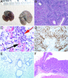
Fig. 1.
Fig. 1.
Pathology at 4 months after transfer of 3 × 10⁴ CD4 T cells into Rag2−/−mice. (A) Gross pathology of lungs from normal Rag2−/−mice and mice that had received 3 × 10⁴ CD4 T cells. (B and C) H&E stain (B) and Luna stain (C) for eosinophils (red arrow) and eosinophilic crystal-laden macrophages (black arrow). (D and E) Immunohistochemical anti-Ym1 stain of lungs from mice that had received 3 × 10⁴ CD4 T cells (D) and of lungs from normal mice (E). (F) H&E stain of junction of forestomach and glandular stomach from mice that had received 3 × 10⁴ CD4 T cells showing eosinophilic and lymphocytic infiltrate with parietal cell loss.
Fig. 2.
Fig. 2.
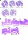
Representative H&E sections of whole lung (A) and junction of glandular stomach and forestomach (B) from Rag2−/−mice 4 months after transfer of 30,000 CD25− CD44lo CD4 T cells, 30,000 CD4 T cells, 2 million CD25− CD44lo CD4 T cells, or 2 million CD4 T cells and from an age-matched Rag2−/− control that had not received cells.
Fig. 3.
Fig. 3.
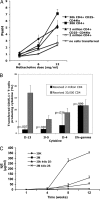
Th2 phenotype predominates in diseased mice. (A) Methacholine hypersensitivity. Mice were placed individually in a whole-body plethysmograph (Buxco Electronics), and Penh values were calculated. There were three to five mice per group. Doses of methacholine >12 mg/ml resulted in death for a number of the mice in the groups receiving 30,000 T cells. (B) Peripheral lymph node cells from Rag2−/− mice that had received 3 × 10⁴ or 2 × 10⁶ CD4 T cells 4 months earlier were stimulated with PMA and ionomycin for 6 h; monensin was added for the last 2 h. Cells were stained for CD45.1 to identify transferred cells. Cells were then fixed, permeabilized, and stained for intracellular cytokines. Each group consisted of four to five mice; shown is a representative experiment of three similar experiments. (C) Elevated serum IgE. B10.A CD3ε−/−mice intravenously received the cells indicated and were bled at the times indicated, and serum IgE was measured by ELISA. There were four mice in each group; this experiment was repeated once with similar results. Standard error bars are shown.
Fig. 4.
Fig. 4.
Antiparietal cell antibodies in mice receiving 30,000 CD4 T cells. Fluorescence microscopy to detect antiparietal cell antibodies in serum from CD3ε−/−recipient mice 12 weeks after transfer of 30,000 (Upper Left) or 2 million (Upper Right) sorted CD44lo CD25− CD4 T cells. Mouse serum was incubated on normal mouse stomach sections, and a FITC-labeled F(ab′)2 goat anti-mouse IgG antibody was used for detection. Results from the staining experiment are summarized in Lower.
Fig. 5.
Fig. 5.
Transfer of large but not small numbers of Tregs controls disease. CD45.2 Rag2−/−mice received either 3 × 10⁴ or 3 × 10⁵ sorted CD25+ CD45.1 CD4 T cells intravenously. (A) Lymph nodes and spleens were harvested, and transferred CD45.1 cells were counted 12 weeks later. (B) Ten weeks after primary CD45.2, CD25+ CD4 T cell transfer, 1 million CFSE-labeled CD45.1 CD25− CD4 T cells were transferred, and lymph nodes were harvested 1 week later. Representative CFSE profiles of transferred CD45.1 T cells are shown. (C) Ten weeks after primary CD45.2+ Treg transfer, mice received a second transfer of 1 × 10⁵ CD25− CD45.1+ CD4 T cells. Cells were harvested 10 weeks later. CD45.1+ and CD45.2+ CD4 T cells were enumerated, and pathology of mice initially receiving 3 × 10⁴ Tregs (D and F) or 3 × 10⁵ Tregs (E and G) was examined. Cell yields and representative pathology are shown. There were two to three mice in each group. This experiment was repeated once with similar results. Standard error bars are shown.

Similar articles

Cited by

References

    1. Markert ML, Alexieff MJ, Li J, Sarzotti M, Ozaki DA, Devlin BH, Sempowski GD, Rhein ME, Szabolcs P, Hale LP, et al. J Allergy Clin Immunol. 2000;113:734–741. - PubMed
    1. Villa A, Santagata S, Bozzi F, Giliani S, Frattini A, Imberti L, Gatta LB, Ochs HD, Schwarz K, Notarangelo LD, et al. Cell. 1998;93:885–896. - PubMed
    1. Tietz A, Sponagel L, Erb P, Bucher H, Battegay M, Zimmerli W. Eur J Clin Microbiol Infect Dis. 1997;16:675–677. - PubMed
    1. Yamanaka K, Yawalkar N, Jones DA, Hurwitz D, Ferenczi K, Eapen S, Kupper TS. Clin Cancer Res. 2005;11:5748–5755. - PubMed
    1. Yawalkar N, Ferenczi K, Jones DA, Yamanaka K, Suh KY, Sadat S, Kupper TS. Blood. 2003;102:4059–4066. - PubMed

Publication types

MeSH terms

LinkOut - more resources