. 2007 Jan 9;104(2):606-11.
doi: 10.1073/pnas.0605957104. Epub 2007 Jan 3.

Postnatal lymphatic partitioning from the blood vasculature in the small intestine requires fasting-induced adipose factor

Affiliations

Postnatal lymphatic partitioning from the blood vasculature in the small intestine requires fasting-induced adipose factor

Fredrik Bäckhed et al. Proc Natl Acad Sci U S A. .

Abstract

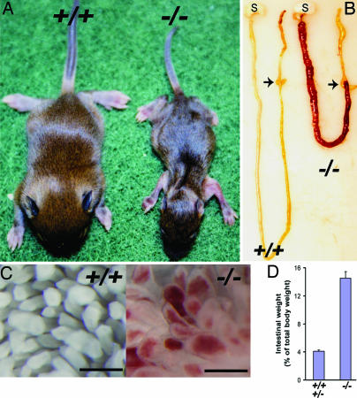
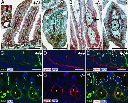
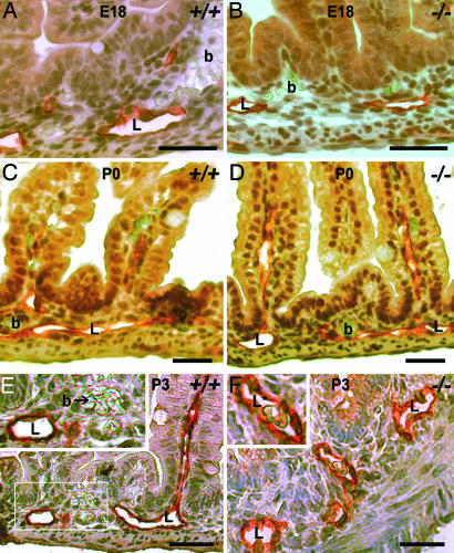
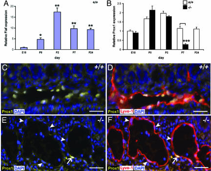
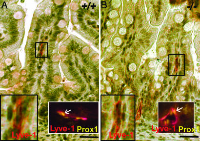
Lymphatic vessels develop from specialized venous endothelial cells. Using knockout mice, we found that fasting-induced adipose factor (Fiaf) is required for functional partitioning of postnatal intestinal lymphatic and blood vessels. In wild-type animals, levels of intestinal Fiaf expression rise during the first postnatal day and peak at day 2, which coincides with the onset of the lymphatico-venous partitioning abnormality in Fiaf-/- mutants on a mixed 129/SvJ:C57BL/6 genetic background. Fiaf deficiency is not associated with disruption of the blood vasculature or with lymphatic endothelial recruitment of smooth muscle cells. We identified Prox1, a critical regulator of lymphangiogenesis, as a downstream target for Fiaf signaling in the intestinal lymphatic endothelium. This organ-specific lymphovascular abnormality can be rescued by allowing embryonic Fiaf-/- intestinal isografts to develop in Fiaf+/+ recipients.


Conflict of interest statement

The authors declare no conflict of interest.

Figures

Fig. 1.
Fig. 1.
Fiaf-deficient mice have blood-filled small intestinal villi. (A) A P12 Fiaf−/− mouse (mixed 129/SvJ:B6 genetic background) exhibits cachexia and a protuberant abdomen compared with its Fiaf+/+ littermate. (B) Small intestine of the P12 Fiaf−/− animal shown in A appears bright red, thickened, and foreshortened. These gross abnormalities terminate abruptly at the ileal–cecal junction (arrows). S, stomach. (C) Whole-mount preparation of small intestines from the mice in A, demonstrating blood-filled Fiaf−/− villi. (Scale bars: 1 mm.) (D) Increased small intestine-to-body weight ratio in P6–P10 Fiaf−/− mice (n = 11) compared with Fiaf+/+ and Fiaf+/− littermates (n = 20) (P < 0.001). Mean values ± SE are plotted.
Fig. 2.
Fig. 2.
Functionally conjoined lymphatic and blood vasculature in the small intestines of Fiaf−/− mice. (A and B) Bright-field immunohistochemistry showing Lyve-1 (red) in hematoxylin-stained sections of small intestine from P7 Fiaf+/+ (A) and Fiaf−/− (B) littermates. Cross-sections of villi from Fiaf+/+ and Fiaf−/− small intestine are displayed in A Right and B Right, respectively. The dilated lymphatic vasculature in the villus core mesenchyme of the Fiaf-deficient mouse is lined by Lyve-1-positive endothelial cells (e.g., arrow) and filled with blood cells (asterisks). (C–H) Immunostaining for Lyve-1 (red) in the small intestines of P7 Fiaf+/+ and Fiaf−/− mice that received an i.v. injection of FITC-labeled BS-1 (blood and lymphatic vascular endothelial cell-binding lectin; green) 30 min before they were killed. Note the presence of dilated FITC-BS-1/Lyve-1-positive lymphatic vessels (orange) at the base of Fiaf−/− villi. Dashed lines in E and H outline the margins of villi. (Scale bars: 50 μm in A and B and 100 μm in C–H.)
Fig. 3.
Fig. 3.
Development of small intestinal lymphatics in wild-type and Fiaf−/− mice. (A–D) Close approximation of anatomically distinct Lyve-1-positive lymphovasculature (L) and blood vasculature (b) in E18 and P0 wild-type (A and C) and Fiaf−/− (B and D) littermates. At these stages of development, no erythrocytes are seen in the lymphovasculature of knockout (or wild-type) littermates. (E and F) By P3, blood cells are present within the lymphatic vasculature (L*), but not in the extravascular space, of Fiaf−/− animals (compare Insets). (Scale bars: 50 μm in A, B, E, and F and 25 μm in C and D.)
Fig. 4.
Fig. 4.
Prox1 expression is regulated in the small intestine by Fiaf. (A) qRT-PCR analysis of the relative expression of Fiaf in the small intestine of wild-type E18–P24 mice (n = 4 per group, each intestine analyzed individually; mean values ± SE are plotted relative to E18). ∗, P < 0.05; ∗∗, P < 0.01. Control experiments showed that Fiaf expression remains constant in the liver during this period (data not shown). (B) Expression of Prox1 in the small intestines of Fiaf+/+ and Fiaf−/− littermates (n = 4 per group, each intestine analyzed individually; mean values ± SE are plotted relative to E18; note that Fiaf−/− mice do not survive to P24, so no data are shown for this time point). ∗∗∗, P < 0.001 compared with wild type. (C–F) Multilabel immunofluorescence study demonstrating Prox1-positive nuclei (yellow) in Lyve-1-positive small intestinal lymphovascular endothelium (red). In wild-type mice (C and D), nearly all lymphatic endothelial cell nuclei are Prox1-positive, whereas in Fiaf−/− mice (E and F), >90% are Prox1-negative (arrowheads). The arrows in E and F point to a rare Prox1-positive lymphatic endothelial cell nucleus within the Fiaf−/− small intestine. (Scale bars: 50 μm.)
Fig. 5.
Fig. 5.
Circulating Fiaf rescues the conjoined lymphatico-venous abnormality in Fiaf−/− isografts. Small intestinal isografts were harvested from E15 Fiaf+/+ or Fiaf−/− donors and implanted for 14 days into the dorsal s.c. tissue of adult Fiaf+/+ recipients. Serial sections of harvested isografts were stained with antibodies to Lyve-1 (red) or Prox1 (yellow) and counterstained with hematoxylin. Representative nascent crypt villus units are shown. Lymphovascular development is indistinguishable in wild-type (A) and Fiaf−/− (B) isografts. Black-bordered insets show enlarged views of the black boxed areas: note the absence of dilated, blood-filled lymphatic vessels in the Fiaf−/− isograft. White-bordered insets show Lyve-1-positive lymphatic endothelium with Prox1-positive nuclei (arrows). (Scale bars: 25 μm.)

Similar articles

Cited by

References

    1. Wigle JT, Oliver G. Cell. 1999;98:769–778. - PubMed
    1. Karkkainen MJ, Haiko P, Sainio K, Partanen J, Taipale J, Petrova TV, Jeltsch M, Jackson DG, Talikka M, Rauvala H, et al. Nat Immunol. 2004;5:74–80. - PubMed
    1. Dumont DJ, Jussila L, Taipale J, Lymboussaki A, Mustonen T, Pajusola K, Breitman M, Alitalo K. Science. 1998;282:946–949. - PubMed
    1. Karkkainen MJ, Ferrell RE, Lawrence EC, Kimak MA, Levinson KL, McTigue MA, Alitalo K, Finegold DN. Nat Genet. 2000;25:153–159. - PubMed
    1. Karkkainen MJ, Saaristo A, Jussila L, Karila KA, Lawrence EC, Pajusola K, Bueler H, Eichmann A, Kauppinen R, Kettunen MI, et al. Proc Natl Acad Sci USA. 2001;98:12677–12682. - PMC - PubMed

Publication types

MeSH terms

LinkOut - more resources