. 2007 May 1;109(9):3794-802.
doi: 10.1182/blood-2005-09-010116. Epub 2007 Jan 3.

An essential role for IL-17 in preventing pathogen-initiated bone destruction: recruitment of neutrophils to inflamed bone requires IL-17 receptor-dependent signals

Affiliations

An essential role for IL-17 in preventing pathogen-initiated bone destruction: recruitment of neutrophils to inflamed bone requires IL-17 receptor-dependent signals

Jeffrey J Yu et al. Blood. .

Abstract

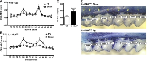
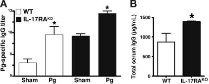
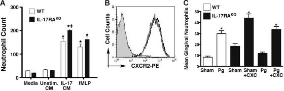
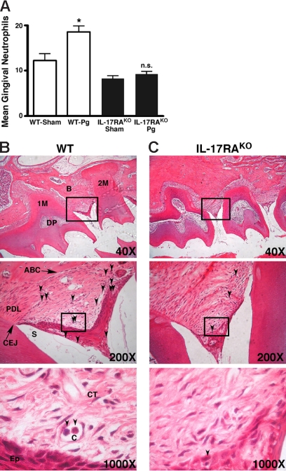
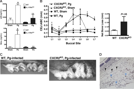
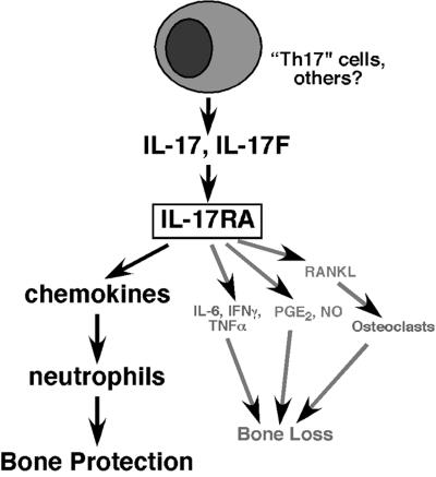
IL-17 and its receptor are founding members of a novel family of inflammatory cytokines. IL-17 plays a pathogenic role in rheumatoid arthritis (RA)-associated bone destruction. However, IL-17 is also an important regulator of host defense through granulopoiesis and neutrophil trafficking. Therefore, the role of IL-17 in pathogen-initiated bone loss was not obvious. The most common form of infection-induced bone destruction occurs in periodontal disease (PD). In addition to causing significant morbidity, PD is a risk factor for atherosclerotic heart disease and chronic obstructive pulmonary disease (COPD). Similar to RA, bone destruction in PD is caused by the immune response. However, neutrophils provide critical antimicrobial defense against periodontal organisms. Since IL-17 is bone destructive in RA but a key regulator of neutrophils, we examined its role in inflammatory bone loss induced by the oral pathogen Porphyromonas gingivalis in IL-17RA-deficient mice. These mice showed enhanced periodontal bone destruction, suggesting a bone-protective role for IL-17, reminiscent of a neutrophil deficiency. Although IL-17RA-deficient neutrophils functioned normally ex vivo, IL-17RA knock-out (IL-17RA(KO)) mice exhibited reduced serum chemokine levels and concomitantly reduced neutrophil migration to bone. Consistently, CXCR2(KO) mice were highly susceptible to alveolar bone loss; interestingly, these mice also suggested a role for chemokines in maintaining normal bone homeostasis. These results indicate a nonredundant role for IL-17 in mediating host defense via neutrophil mobilization.


Figures

Figure 1
Figure 1
IL-17RAKO mice exhibit enhanced alveolar bone loss in response to P gingivalis infection. (A-B) WT and IL-17RAKO mice (n = 6-8) were infected with Pg or sham-infected. Alveolar bone destruction was assessed after 6 weeks by measuring the distance from the ABC to the CEJ at 14 maxillary buccal sites per mouse (R1-R7 = right jaw; L1-L7 = left jaw). Standard deviations are shown. Data were analyzed by a Mann-Whitney unpaired t test, and statistically significant differences of Pg-infected compared with sham-infected for each buccal site are indicated with an asterisk (P < .05). (C) Net bone loss averaged over all buccal sites for WT and IL-17RAKO mice, with standard deviations. (D) Representative bone loss measurements from IL-17RAKO sham-infected (C) and Pg-infected (D) maxillary jaws that were stained with methylene blue. Images were acquired with a Nikon SMZ 1000 microscope, magnification ×3. Buccal sites (1-7) are indicated in yellow, and specific ABC/CEJ distance measurements are indicated in boxes.
Figure 2
Figure 2
IgG titers are elevated in IL-17RAKO mice. (A) Sera from mice described in Figure 1 were analyzed for Pg-specific IgG in WT (□) and IL-17RAKO (■) mice by ELISA. Standard deviations are shown. *Statistically significant differences in Pg-infected samples compared with sham-infected samples of the same strain, as determined by unpaired t test (P < .05). There was no significant difference between WT Pg-infected IgG and IL-17RAKO sham-infected IgG levels. (B) Total IgG levels in uninfected WT (□) and IL-17RAKO (■) mice were determined in triplicate by ELISA. Standard deviations are shown. *Statistically significant difference compared with WT determined by unpaired t test (P < .05).
Figure 3
Figure 3
Characterization of IL-17RAKO neutrophils. (A) IL-17RAKO neutrophils show no migration defects ex vivo. As a source of chemokines, conditioned media (CM) was obtained from MC3T3-E1 cells stimulated with nothing (Unstim. CM) or 200 ng/mL IL-17 (IL-17 CM; contains > 39 ng/mL LIX; data not shown) for 24 hours (as described). Bone marrow–derived Gr1+ cells from WT (□) or IL-17RAKO mice (■) were incubated with media alone, with MC3T3-E1 CM, or with the chemotactic peptide fMLP (100 μM). Cells in the lower chambers were quantified in 10 random microscope fields, and standard deviations are shown. *Significant differences compared with unstimulated samples (P < .001); ‡significant difference between WT and IL-17RAKO mice for the same treatment condition (P < .01). (B) Normal CXCR2 levels in IL-17RAKO mice. Bone marrow from WT (black) or IL-17RAKO (gray) mice was stained with Abs to Gr1 and CXCR2. Cells were gated on the Gr1+ population, and the profile of CXCR2 staining is shown compared with the isotype control. (C) Ectopically applied chemokines rescue neutrophil migration to gingiva in IL-17RAKO mice. WT and IL-17RAKO mice were anesthetized, and a mixture of LIX and Groα was applied adjacent to the maxillary first molars for 1 hour. Sagittal sections of maxillary tissue were stained with H&E, and neutrophils were assessed by counting in a randomized, blinded fashion. Standard deviations are shown. *Significant differences compared with sham-infected sample by unpaired t test (P < .05).
Figure 4
Figure 4
Defective neutrophil migration to the gingiva in IL-17RAKO mice. IL-17RAKO or WT mice were infected with Pg 3 times over 5 days and killed 24 hours after the final inoculation. Sections were stained with H&E. Images were acquired with a Zeiss Axioimager 2I microscope system. (A) At least 80 slides per condition were examined for neutrophils by oil immersion microscopy at ×1000 magnification, and the number of neutrophils normalized to number of teeth present per slide was determined. Standard deviations are shown. Statistical significance of differences between Pg-infected and sham-infected was assessed by unpaired t test (P < .05). (B-C) Sample slides showing neutrophils in representative Pg-infected WT (panel B) or IL-17RAKO (panel C) gingival slices imaged with 40×/0.10, 200×/0.45, and 1000×/1.30 oil-immersion objective lenses. Arrows indicate polymorphonuclear neutrophils. 1M indicates first molar; 2M, second molar; PDL, periodontal ligament; B, bone; ABC, alveolar bone crest; CEJ, cementoenamel junction; DP, dental pulp; CT, connective tissue; Ep, epithelium; C, capillary; and S, sulcus.
Figure 5
Figure 5
CXC chemokine expression in situ. (A) IL-17RAKO mice fail to up-regulate CXC chemokines in response to Pg infection. Serum samples from WT and IL-17RAKO mice in Figure 1 were analyzed for Groα and LIX by ELISA. Standard deviations are shown. *Statistically significant differences between Pg-infected compared with Sham-infected mouse strains as assessed by unpaired t test (LIX, P < .001; Groα, P < .05). n.s. indicate not significant. (B) CXCR2KO mice are susceptible to Pg-induced bone loss. CXCR2KO or WT mice (BALB/c background) were infected with Pg or sham (n = 4-6), and ABC/CEJ distances on the left maxillary jaw were evaluated as in Figure 1. Net bone loss with standard deviations is shown. (C) Representative images of maxillary jaws in Pg-infected WT or CXCR2KO mice, taken as in Figure 1D. (D) Source of CXC chemokines in Pg infection. Sections from Pg-infected WT mice (adjacent sections as in Figure 4B) were stained with antisera that recognizes Groα, LIX, and MIP2. Blue arrows indicate representative monocyte/macrophages; black arrows indicate representative fibroblasts. Image obtained with a 200×/0.45 objective lens. n.s. indicates not significant.
Figure 6
Figure 6
Opposing roles of IL-17 in inflammatory bone loss. While the dominant influence of IL-17 in RA leads to bone destruction (see Moseley et al, Gaffen, and Lubberts et al), we find the net effect of IL-17RA–mediated signaling in PD leads to alveolar bone protection. IL-17 is produced primarily by T cells, and triggers a variety of target cells to secrete inflammatory mediators, including chemokines, cytokines, cell-surface receptors, prostaglandin E2, and nitric oxide (NO). While many of these effectors exert bone resorptive effects by mediating enhanced osteoclastogenesis, chemokines and the neutrophils they recruit exert antimicrobial activities that ultimately lead to a bone-protective effect in the context of periodontal infection.

Similar articles

Cited by

References

    1. Moseley TA, Haudenschild DR, Rose L, Reddi AH. Interleukin-17 family and IL-17 receptors. Cytokine Growth Factor Rev. 2003;14:155–174. - PubMed
    1. Kelly MN, Kolls JK, Happel K, et al. Interleukin-17/interleukin-17 receptor-mediated signaling is important for generation of an optimal polymorphonuclear response against Toxoplasma gondii infection. Infect Immun. 2005;73:617–621. - PMC - PubMed
    1. Ye P, Rodriguez FH, Kanaly S, et al. Requirement of interleukin 17 receptor signaling for lung CXC chemokine and granulocyte colony-stimulating factor expression, neutrophil recruitment, and host defense. J Exp Med. 2001;194:519–527. - PMC - PubMed
    1. Huang W, Na L, Fidel PL, Schwarzenberger P. Requirement of interleukin-17A for systemic anti-Candida albicans host defense in mice. J Infect Dis. 2004;190:624–631. - PubMed
    1. Langrish CL, Chen Y, Blumenschein WM, et al. IL-23 drives a pathogenic T cell population that induces autoimmune inflammation. J Exp Med. 2005;201:233–240. - PMC - PubMed

Publication types

MeSH terms