

Human C-reactive protein protects mice from Streptococcus pneumoniae infection without binding to pneumococcal C-polysaccharide
- PMID: 17202380
- PMCID: PMC3818096
- DOI: 10.4049/jimmunol.178.2.1158
Human C-reactive protein protects mice from Streptococcus pneumoniae infection without binding to pneumococcal C-polysaccharide
Abstract
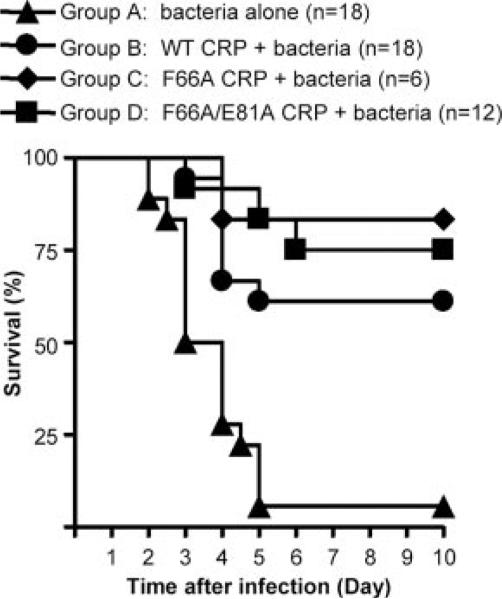
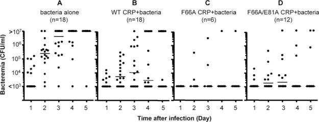
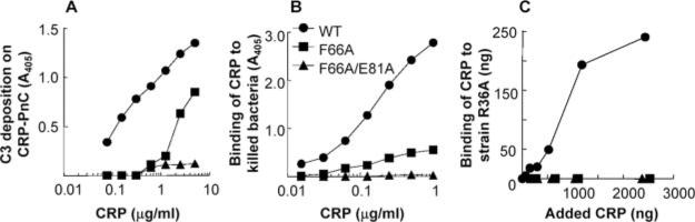
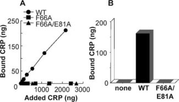
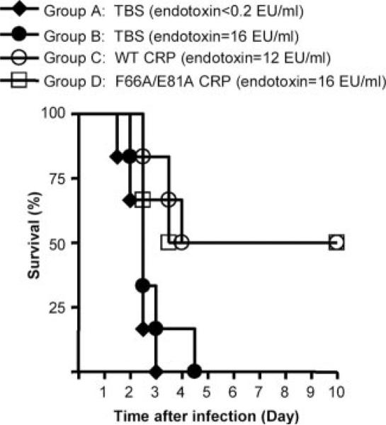
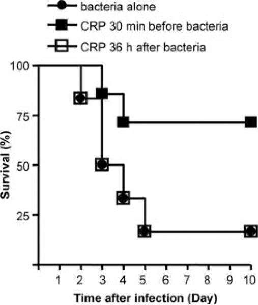
Human C-reactive protein (CRP) protects mice from lethality after infection with virulent Streptococcus pneumoniae type 3. For CRP-mediated protection, the complement system is required; however, the role of complement activation by CRP in the protection is not defined. Based on the in vitro properties of CRP, it has been assumed that protection of mice begins with the binding of CRP to pneumococcal C-polysaccharide on S. pneumoniae and subsequent activation of the mouse complement system. In this study, we explored the mechanism of CRP-mediated protection by utilizing two CRP mutants, F66A and F66A/E81A. Both mutants, unlike wild-type CRP, do not bind live virulent S. pneumoniae. We found that passively administered mutant CRP protected mice from infection as effectively as the wild-type CRP did. Infected mice injected with wild-type CRP or with mutant CRP lived longer and had lower mortality than mice that did not receive CRP. Extended survival was caused by the persistence of reduced bacteremia in mice treated with any CRP. We conclude that the CRP-mediated decrease in bacteremia and the resulting protection of mice are independent of an interaction between CRP and the pathogen and therefore are independent of the ability of CRP to activate mouse complement. It has been shown previously that the Fcgamma receptors also do not contribute to such CRP-mediated protection. Combined data lead to the speculation that CRP acts on the effector cells of the immune system to enhance cell-mediated cytotoxicity and suggest investigation into the possibility of using CRP-loaded APC-based strategy to treat microbial infections.
Figures
Similar articles
-
Role of the property of C-reactive protein to activate the classical pathway of complement in protecting mice from pneumococcal infection.J Immunol. 2006 Apr 1;176(7):4369-74. doi: 10.4049/jimmunol.176.7.4369. J Immunol. 2006. PMID: 16547275 Free PMC article.
-
Protection from Streptococcus pneumoniae infection by C-reactive protein and natural antibody requires complement but not Fc gamma receptors.J Immunol. 2002 Jun 15;168(12):6375-81. doi: 10.4049/jimmunol.168.12.6375. J Immunol. 2002. PMID: 12055255
-
Protection against prolonged pneumococcal infection involves structural changes in C-reactive protein and subsequent binding to both phosphocholine and amyloids on the bacterial surface.Front Immunol. 2025 Jul 16;16:1631409. doi: 10.3389/fimmu.2025.1631409. eCollection 2025. Front Immunol. 2025. PMID: 40740789 Free PMC article.
-
The protective function of human C-reactive protein in mouse models of Streptococcus pneumoniae infection.Endocr Metab Immune Disord Drug Targets. 2008 Dec;8(4):231-7. doi: 10.2174/187153008786848321. Endocr Metab Immune Disord Drug Targets. 2008. PMID: 19075776 Free PMC article. Review.
-
Structure-Function Relationships of C-Reactive Protein in Bacterial Infection.Front Immunol. 2019 Feb 26;10:166. doi: 10.3389/fimmu.2019.00166. eCollection 2019. Front Immunol. 2019. PMID: 30863393 Free PMC article. Review.
Cited by
-
Deficiency of C-reactive protein or human C-reactive protein transgenic treatment aggravates influenza A infection in mice.Front Immunol. 2022 Oct 6;13:1028458. doi: 10.3389/fimmu.2022.1028458. eCollection 2022. Front Immunol. 2022. PMID: 36275680 Free PMC article.
-
Identification of acidic pH-dependent ligands of pentameric C-reactive protein.J Biol Chem. 2010 Nov 12;285(46):36235-44. doi: 10.1074/jbc.M110.142026. Epub 2010 Sep 14. J Biol Chem. 2010. PMID: 20843812 Free PMC article.
-
Predictors of treatment failure and clinical stability in patients with community acquired pneumonia.Ann Transl Med. 2017 Nov;5(22):443. doi: 10.21037/atm.2017.06.54. Ann Transl Med. 2017. PMID: 29264360 Free PMC article. Review.
-
C-Reactive Protein-Based Strategy to Reduce Antibiotic Dosing for the Treatment of Pneumococcal Infection.Front Immunol. 2021 Jan 20;11:620784. doi: 10.3389/fimmu.2020.620784. eCollection 2020. Front Immunol. 2021. PMID: 33552084 Free PMC article.
-
Low C-reactive protein values at admission predict mortality in patients with severe community-acquired pneumonia caused by Streptococcus pneumoniae that require intensive care management.Infection. 2015 Apr;43(2):193-9. doi: 10.1007/s15010-015-0755-0. Epub 2015 Mar 3. Infection. 2015. PMID: 25732200 Free PMC article.
References
-
- Kushner I. The phenomenon of the acute phase response. Ann. NY Acad. Sci. 1982;389:39–48. - PubMed
-
- Volanakis JE. Human C-reactive protein: expression, structure, and function. Mol. Immunol. 2001;38:189–197. - PubMed
-
- Volanakis JE, Kaplan MH. Specificity of C-reactive protein for choline phosphate residues of pneumococcal C-polysaccharide. Proc. Soc. Exp. Biol. Med. 1971;136:612–614. - PubMed
-
- Kaplan MH, Volanakis JE. Interaction of C-reactive protein complexes with the complement system. I. Consumption of human complement associated with the reaction of C-reactive protein with pneumococcal C-polysaccharide and with the choline phosphatides, lecithin, and sphingomyelin. J. Immunol. 1974;112:2135–2147. - PubMed
Publication types
MeSH terms
Substances
Grants and funding
LinkOut - more resources
Full Text Sources
Medical
Research Materials
Miscellaneous
