. 2007 Jan 3;14(1-2):63-74.
doi: 10.1101/lm.439007. Print 2007 Jan-Feb.

Opposing actions of chronic Delta9-tetrahydrocannabinol and cannabinoid antagonists on hippocampal long-term potentiation

Affiliations

Opposing actions of chronic Delta9-tetrahydrocannabinol and cannabinoid antagonists on hippocampal long-term potentiation

Alexander F Hoffman et al. Learn Mem. .

Abstract

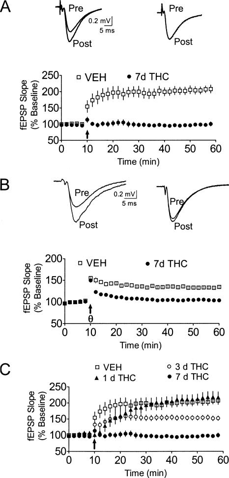
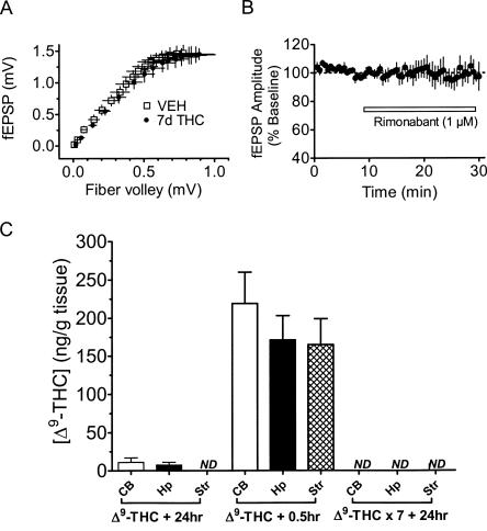
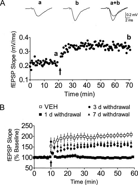
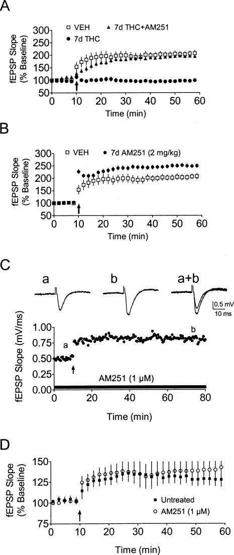
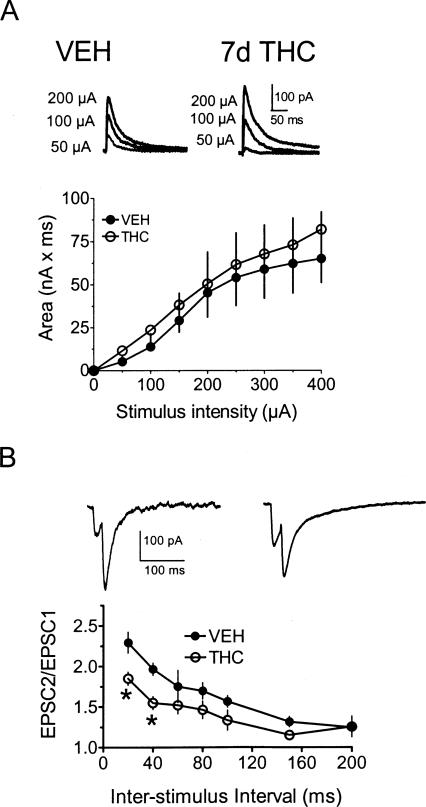
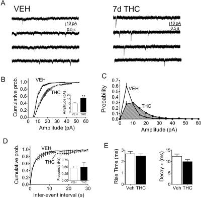
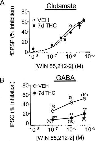
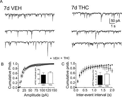
Memory deficits produced by marijuana arise partly via interaction of the psychoactive component, Delta(9)-tetrahydrocannabinol (Delta(9)-THC), with cannabinoid receptors in the hippocampus. Although cannabinoids acutely reduce glutamate release and block hippocampal long-term potentiation (LTP), a potential substrate for learning and memory, the consequences of prolonged exposure to Delta(9)-THC for hippocampal function are poorly understood. Rats were injected with Delta(9)-THC (10 mg/kg, i.p., q.d.) for 1, 3, or 7 d, and electrophysiological recordings were performed in hippocampal slices 1d after the final injection. At this time, Delta(9)-THC was undetectable in hippocampus using liquid chromatography-mass spectrometry (LC-MS). Hippocampal LTP generated using high-frequency (HFS) or theta burst stimulation was not observed in brain slices from the 7-d Delta(9)-THC-treated animals. Delta(9)-THC also blocked HFS-LTP after 3 d, but not 1 d of treatment. The complete blockade of LTP persisted for 3 d after the last Delta(9)-THC injection, and full reversal of the LTP deficit was not observed up to 14 d following Delta(9)-THC withdrawal. The cannabinoid antagonist AM251 (2 mg/kg), administered before each Delta(9)-THC injection prevented the blockade of LTP, and 7-d treatment with AM251 alone significantly increased the level of LTP. Chronic Delta(9)-THC also produced tolerance to the inhibition of synaptic GABA, but not glutamate release by the agonist WIN55,212-2. These data define consequences of repeated Delta(9)-THC exposure for synaptic plasticity in the hippocampus that may help explain memory impairments in humans following chronic marijuana use.


Figures

Figure 1.
Figure 1.
Blockade of LTP following repeated daily treatment with Δ9-THC. (A, top panel) Averaged (5 sweeps) fEPSPs obtained prior to (Pre) and 60 min following (Post) delivery of high-frequency LTP-inducing stimulation (HFS) recorded in hippocampal brain slices 24 h after a 7-d treatment with either vehicle (left) or 10 mg/kg Δ9-THC (right). (Bottom panel) Mean time course of fEPSP slope measured in brain slices obtained from vehicle (VEH) and chronic Δ9-THC-treated rats. The arrow indicates the timing of HFS. (B) Blockade of LTP induced by theta-burst stimulation (TBS, θ). In slices from vehicle-treated rats, TBS-LTP persisted for >60 min following stimulation. TBS-LTP was not observed in slices from Δ9-THC-treated rats. (C) Mean time course of HFS-LTP observed in brain slices obtained from animals treated with vehicle, or with Δ9-THC for 1, 3, or 7 d.
Figure 2.
Figure 2.
The blockade of HFS-LTP measured at 24 h after 7-d treatment with chronic Δ9-THC does not result from the accumulation of the drug in brain tissue, nor from altered brain slice viability. (A) Plot of input–output relationship of presynaptic fiber volley to fEPSP amplitude in hippocampal slices from 7-d vehicle (VEH) and 7-d Δ9-THC-treated (THC) rats. No differences were observed between the two groups. (B) Bath application of the CB1R antagonist rimonabant (SR141716A) did not alter fEPSPs in slices from 7-d Δ9-THC-treated rats, suggesting that receptors were not tonically activated by the drug. (C) Detection of Δ9-THC in rodent brain tissue using LC-MS, at varying times after its acute or chronic injection. Δ9-THC levels were measured in the cerebellum (CB), hippocampus (Hp), or striatum (Str) at either 0.5 or 24 h following a single injection of the drug (Δ9-THC + 0.5hr, Δ9-THC + 24hr, respectively), or 24 h following a once daily, 7-d treatment with the drug (Δ9-THC × 7 + 24hr). Meaningful concentrations of Δ9-THC were only measured in brain tissue from animals in the Δ9-THC + 0.5 h group, suggesting that at the time point used in most of our physiological studies of LTP (i.e., Δ9-THC × 7 + 24hr), the drug was no longer present at pharmacologically relevant concentrations in the hippocampus. (ND) Nondetectable.
Figure 3.
Figure 3.
Partial recovery of HFS-LTP during withdrawal from 7 d of treatment with Δ9-THC. (A) A single experiment that was conducted 7 d following a 7-d exposure to Δ9-THC. The averaged fEPSP waveforms (n = 5 sweeps) were obtained at the points indicated in the time course below. The arrow indicates the timing of HFS. Note the robust HFS-LTP seen in this representative example. (B) Summary of HFS-LTP recovery in hippocampal slices following either 1 d, 3 d, or 7 d of withdrawal from Δ9-THC. The level of HFS-LTP obtained in the 3-d, 7-d, and 14-d (data not shown) withdrawal groups all differed significantly from both the 1-d and the vehicle-injected control.
Figure 4.
Figure 4.
The CB1R antagonist AM251 reverses the blockade of HFS-LTP by Δ9-THC, and increases LTP when administered chronically. (A) Mean effect of chronic treatment (7 d) with Δ9-THC (10 mg/kg), Δ9-THC + AM251, or vehicle on HFS-LTP (arrow) in hippocampal slices. AM251 (2 mg/kg) was administered 30 min prior to each of seven daily Δ9-THC injections. (B) Chronic AM251 enhancement of HFS-LTP. Animals were given i.p. injections of AM251, alone, for 7 d (2 mg/kg). Hippocampal HFS-LTP was measured 24 h following the last AM251 injection. HFS-LTP was significantly enhanced compared to vehicle-treated controls. (C) Time course from a single hippocampal slice experiment in which HFS-LTP was induced during acute AM251 (1 μM) treatment. HFS was delivered at the time indicated by the arrow, and averaged fEPSPs were obtained at the indicated times. (D) Mean effects of acute AM251 exposure on HFS-LTP. No significant differences in HFS-LTP were observed between untreated and AM251-treated slices obtained from drug-naive animals.
Figure 5.
Figure 5.
Chronic exposure to Δ9-THC does not alter NMDA receptor function, but reduces paired pulse facilitation of AMPA-mediated EPSCs. (A) Input–output relationship between stimulus intensity and whole-cell NMDA-mediated synaptic EPSC charge (area = nA × msec) recorded from CA1 pyramidal cells in brain slices obtained from animals treated with Δ9-THC or vehicle for 7 d. Outward NMDA currents were elicited via electrical stimulation of the sc fibers in neurons voltage-clamped at +40 mV, during application of picrotoxin (100 μM) and DNQX (40 μM). Representative mean traces of NMDA currents at varying stimulus intensities are shown for both a vehicle-treated and a Δ9-THC-treated neuron. No significant differences were observed. (B) Paired-pulse facilitation of evoked AMPA-mediated EPSCs at varying interstimulus intervals. Representative traces from slices obtained from a vehicle-treated (left) and THC-treated (right) rat, using a 20-msec paired pulse interval. Synaptic facilitation was significantly reduced in THC-treated slices at shorter interpulse intervals, relative to vehicle controls.
Figure 6.
Figure 6.
Analysis of spontaneous EPSCs (sEPSCs) in hippocampal CA1 pyramidal neurons from vehicle and 7-d Δ9-THC-treated rats. (A) Sample traces of sEPSCs obtained in whole-cell recordings (Vhold −70 mV) single neurons under each condition. Note the larger amplitude of events in the traces on the right. (B) Mean cumulative probability plot and mean amplitude from all neurons (n = 13 neurons/8 vehicle-treated rats; n = 15 neurons/9 rats Δ9-THC-treated animals). A significant increase in the mean sEPSC amplitude was observed during Δ9-THC withdrawal. (C) Mean probability distribution of sEPSCs obtained in each group of neurons. Note the rightward shift in the sEPSC amplitude distribution, and the decrease in smaller sEPSCs (<∼7 pA) in the neurons obtained from the Δ9-THC-treated group. (D) Mean cumulative interevent interval distribution and mean frequency of sEPSCs from the same cells. No differences in sEPSC frequency were observed. (E) Mean rise and decay times of sEPSCs. No significant differences were observed in either parameter.
Figure 7.
Figure 7.
Differential tolerance to WIN 55,212-2 at GABAergic and glutamatergic synapses following withdrawal from repeated Δ9-THC. (A) Concentration-response curves for the effect of WIN55,212-2 on fEPSPs in brain slices recorded 24 h after a 7-d treatment with either Δ9-THC or vehicle. No significant differences between groups were observed (vehicle EC50 = 465 nM; Δ9-THC-treated EC50 = 576 nM). (B) Whole-cell recordings of the concentration-dependent effects of WIN55,212-2 on electrically evoked GABAA IPSCs in hippocampal slices obtained from 7-d vehicle- and 7-d Δ9-THC-treated rats. The inhibition of the evoked IPSCs by WIN55,212-2 was significantly (**P < 0.01; ANOVA) reduced at each concentration following Δ9-THC treatment, indicating the development of tolerance. The number of neurons used at each concentration, under each chronic treatment condition, is shown in parentheses. Note that the fEPSP and the evoked IPSC concentration-response curves are plotted on the same abscissa.
Figure 8.
Figure 8.
Effects of chronic Δ9-THC treatment on GABAergic synaptic function. (A) Spontaneous, GABAA IPSCs (sIPSCs) from a 7-d vehicle-treated, and a 7-d Δ9-THC-treated rat were recorded in the presence of ionotropic glutamate receptor antagonists. (B) Mean cumulative probability plot and mean amplitudes of sIPSCs for all neurons recorded under each condition (vehicle: n = 6 neurons/3 rats; Δ9-THC: n = 9 neurons/5 rats). (C) Average cumulative probability plot of the interevent interval and mean frequency of sIPSCs from the same cells. No differences (K-S > 0.05 for cumulative plots, and P > 0.05 for mean values) in either the amplitude or frequency of the events were observed between these two groups, indicating that basal GABAergic synaptic function was unaltered following repeated Δ9-THC.

Similar articles

Cited by

References

    1. Bliss T.V., Collingridge G.L. A synaptic model of memory: Long-term potentiation in the hippocampus. Nature. 1993;361:31–39. - PubMed
    1. Blitzer R.D., Wong T., Nouranifar R., Iyengar R., Landau E.M. Postsynaptic CAMP pathway gates early LTP in hippocampal CA1 region. Neuron. 1995;15:1403–1414. - PubMed
    1. Bohme G.A., Laville M., Ledent C., Parmentier M., Imperato A. Enhanced long-term potentiation in mice lacking cannabinoid CB1 receptors. Neuroscience. 2000;95:5–7. - PubMed
    1. Bolla K.I., Brown K., Eldreth D., Tate K., Cadet J.L. Dose-related neurocognitive effects of marijuana use. Neurology. 2002;59:1337–1343. - PubMed
    1. Boyd S.T., Fremming B.A. Rimonabant—A selective CB1 antagonist. Ann. Pharmacother. 2005;39:684–690. - PubMed

Publication types

MeSH terms

LinkOut - more resources