. 2007 Jan 3;27(1):46-58.
doi: 10.1523/JNEUROSCI.3966-06.2007.

Prevention of plasticity of endocannabinoid signaling inhibits persistent limbic hyperexcitability caused by developmental seizures

Affiliations

Prevention of plasticity of endocannabinoid signaling inhibits persistent limbic hyperexcitability caused by developmental seizures

Kang Chen et al. J Neurosci. .

Abstract

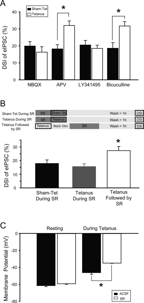
Depolarization-induced suppression of inhibition (DSI) is an endocannabinoid-mediated short-term plasticity mechanism that couples postsynaptic Ca2+ rises to decreased presynaptic GABA release. Whether the gain of this retrograde synaptic mechanism is subject to long-term modulation by glutamatergic excitatory inputs is not known. Here, we demonstrate that activity-dependent long-term DSI potentiation takes place in hippocampal slices after tetanic stimulation of Schaffer collateral synapses. This activity-dependent, long-term plasticity of endocannabinoid signaling was specific to GABAergic synapses, as it occurred without increases in the depolarization-induced suppression of excitation. Induction of tetanus-induced DSI potentiation in vitro required a complex pathway involving AMPA/kainate and metabotropic glutamate receptor as well as CB1 receptor activation. Because DSI potentiation has been suggested to play a role in persistent limbic hyperexcitability after prolonged seizures in the developing brain, we used these mechanistic insights into activity-dependent DSI potentiation to test whether interference with the induction of DSI potentiation prevents seizure-induced long-term hyperexcitability. The results showed that the in vitro, tetanus-induced DSI potentiation was occluded by previous in vivo fever-induced (febrile) seizures, indicating a common pathway. Accordingly, application of CB1 receptor antagonists during febrile seizures in vivo blocked the seizure-induced persistent DSI potentiation, abolished the seizure-induced upregulation of CB1 receptors, and prevented the emergence of long-term limbic hyperexcitability. These results reveal a new form of activity-dependent, long-term plasticity of endocannabinoid signaling at perisomatic GABAergic synapses, and demonstrate that blocking the induction of this plasticity abolishes the long-term effects of prolonged febrile seizures in the developing brain.


Figures

Figure 1.
Figure 1.
Increased neuronal activity leads to potentiation of DSI. A, B, Strong (5 × 10 s at 100 Hz) tetanic stimulation in the stratum radiatum (including the Schaffer collateral pathway) in slices from control animals increased DSI amplitude and prolonged the DSI decay in CA1 pyramidal cells. Example traces are shown in A; summary data are shown in B (based on 3 DSI episodes per cell separated by 1 min). Square waves above traces and summary graph indicate time and duration of depolarization. C, Even after tetanic stimulation, DSI is completely blocked in the presence of the CB1 receptor antagonist SR141716 (1 μm). D, Tetanic stimulation and hyperthermic seizures (performed 1 week before recording), both potentiate DSI to a similar extent. Additionally, tetanic stimulation cannot further potentiate DSI in slices from HT animals, suggesting a common mechanism of potentiation. E, The potentiation of DSI requires a strong tetanic stimulation, as a weaker protocol (5 × 1 s at 100 Hz) does not increase DSI. For all experiments in this figure, DSI was induced by a 500 ms depolarization to 0 mV and recorded in the presence of carbachol and ionotropic glutamate receptor blockers (see Materials and Methods). Recordings were made at least 1 h after sham stimulation (sham-tet) or tetanus (tetanus). In this and following figures, asterisks indicate significant differences (p < 0.05).
Figure 2.
Figure 2.
Tetanic stimulation rapidly potentiates DSI of eIPSCs, increases the sensitivity of eIPSCs to CB1 receptor antagonists/inverse agonists, and does not affect the magnitude of DSE. A, B, Tetanic stimulation potentiates DSI of eIPSCs, and the full effect of potentiation is present within 15 min after stimulation. Representative traces are shown in A, summary data are shown in B. Square wave in B represents time and duration of depolarizing pulse. IPSCs were evoked with stimulation at the border of stratum radiatum and stratum pyramidale. DSI was induced by a 500 ms depolarization to 0 mV and recorded in the presence of ionotropic glutamate receptor blockers, but without carbachol. C, In agreement with the lack of change in sIPSC charge transfer after tetanus, the amplitude of evoked IPSCs is not changed by tetanic stimulation for any stimulation intensity tested (note that the stimulating and recording electrodes were carefully positioned in a reproducible manner) (Chen et al., 1999, 2001) (see Materials and Methods). D, Tetanic stimulation potentiates DSI evoked by either 100 or 500 ms depolarizing pulses. E, Tetanic stimulation causes eIPSCs to become sensitive to the CB1 antagonist SR141716. The inset shows representative traces. Calibration: 50 ms, 100 pA. Symbols refer to E and F. F, The potentiation of endocannabinoid signaling caused by tetanic stimulation is specific to inhibitory synapses, as the amplitude and decay of DSE are not affected by tetanus. DSE was evoked by a 10 s depolarizing pulse and was recorded in the presence of bicuculline (10 μm). The square wave represents the time and duration of the depolarizing pulse. The inset shows representative traces. Calibration: 80 ms, 400 pA.
Figure 3.
Figure 3.
Paired recordings show that DSI at CB1 expressing interneuron to pyramidal cell synapses is potentiated by tetanic stimulation. A, Biocytin fill from a representative interneuron demonstrates the presence of CCK immunostaining and the absence of PV immunostaining (all cells that were successfully recovered were CCK positive and PV negative; n = 9 of 14 recordings). Traces on the right demonstrate the characteristic firing pattern and hyperpolarizing response of CCK-positive interneurons after the injection of +140 pA and − 80pA current pulses. These cells are similar to those examined and illustrated by Foldy et al. (2006). B, DSI of uIPSCs is significantly increased by tetanic stimulation. Top traces show action potentials evoked in the presynaptic basket cell; bottom traces show 20 individual uIPSCs in the postsynaptic pyramidal cell from each time point (gray) as well the averaged uIPSC (black). The time points were as follows, with the zero time point being the start of the depolarizing pulse: baseline or pre-DSI period, −2 s to 0 s; DSI, 0.5 to 2.5 s; recovery, 4 to 6 s. The illustrated traces are consecutive responses obtained by triggering action potentials in the presynaptic interneuron at 10 Hz; as described previously in detail (Foldy et al. 2006), this stimulation frequency resulted in stable responses over time and also allowed us to obtain a sufficiently large number of events for reliable analysis. Calibration: B, presynaptic, 25 mV, 5 ms; postsynaptic, 25 pA, 5 ms. All experiments in this figure were performed in control ACSF and DSI was evoked by 500 ms pulses.
Figure 4.
Figure 4.
Activation of AMPA/kainate, mGluR, and CB1 receptors is required during tetanus for potentiation of DSI. A, Tetanus-induced potentiation of DSI is blocked when NBQX (5 μm), an AMPA/kainate receptor antagonist, or LY341495 (200 μm), a broad spectrum mGluR antagonist, is present during tetanization. Blockade of NMDARs with APV (10 μm), or blockade of GABAA receptors with bicuculline (10 μm) during tetanization does not prevent potentiation of DSI. B, Potentiation of DSI is prevented when SR141716, a CB1R antagonist, is present during tetanus (SR was applied for 6 min, which was followed by a >60 min wash in ACSF). To exclude the possibility that this effect was caused by an incomplete wash out of SR from the slice, a control experiment was performed in which tetanus was applied in the absence of SR, followed by a 6 min perfusion of the slice with SR, followed by a 60 min wash with ACSF (tetanus followed by SR). The presence of potentiated DSI in this experiment indicates that SR can be completely eliminated from the slice with a 60 min wash. The top panel demonstrates experimental protocol, where dark boxes indicate presence of SR in extracellular medium. C, The presence of SR during tetanus does not decrease the effective activation of excitatory afferents. In fact, SR did not change the pretetanus resting membrane potential, but the average membrane potential during tetanic stimulation was depolarized in the presence of SR and, as detailed in the text, the number of action potentials elicited by each tetanic train was also increased in the presence of SR.
Figure 5.
Figure 5.
Blocking CB1 receptors in vivo during experimental febrile seizures prevents both potentiation of DSI and increase in CB1 receptor number. A, The potentiation of DSI observed after HT is prevented when SR is injected in vivo before HT induction at a dose of either 1 or 10 mg/kg. B, Typical western blots of hippocampal tissue (top) and their quantitative analysis (bottom) demonstrate a significant (35.9%) increase in the amount of CB1 receptor protein after HT (HT+vehicle) as compared with littermate controls (either vehicle- or SR-injected controls). The injection of SR in vivo (1 mg/kg) before HT induction (HT+SR) prevents the increased expression of CB1 protein.
Figure 6.
Figure 6.
Blocking CB1 receptors in vivo during febrile seizures prevents the long-lasting decrease in seizure threshold. A–F, Seizure thresholds were tested both with electrical stimulation in vitro at 1 week after febrile seizures (A, B) and with kainate in vivo 6 weeks after febrile seizures (C–F). A, One train of brief high-frequency test stimulation of the Schaffer collaterals (2 s at 60 Hz) in hippocampal-entorhinal slices leads to self-sustaining epileptiform discharges in vehicle-injected HT animals, but not in SR-injected HT animals or in controls. Recordings were performed 1 week after HT induction. Note that the high-frequency stimulus train is used here to test the excitability of the network, not to induce changes in DSI (the test stimulation is considerably shorter than what is required for DSI potentiation). B, Plot of stimulation train number versus the duration of spontaneous field discharges shows that the injection of SR, at 1 mg/kg, prevents the increase in epileptiform activity seen after HT. C, Representative traces from hippocampal depth EEG recordings demonstrate that, 10 min after injection of kainate (5 mg/kg), seizures have initiated in HT+vehicle animals but not in HT + SR (1 mg/kg) animals or control animals. D, The latency to seizure initiation is significantly decreased by HT (HT+vehicle), but this decrease is prevented by injection of SR (HT+SR). E, F, SR injection prevents the increase seen after HT in both the duration of the longest single seizure and the cumulative duration of all seizures.

Similar articles

Cited by

References

    1. Annegers JF, Hauser WA, Shirts SB, Kurland LT. Factors prognostic of unprovoked seizures after febrile convulsions. N Engl J Med. 1987;316:493–498. - PubMed
    1. Baram TZ, Gerth A, Schultz L. Febrile seizures: an appropriate-aged model suitable for long-term studies. Brain Res Dev Brain Res. 1997;98:265–270. - PMC - PubMed
    1. Beau FE, Alger BE. Transient suppression of GABAA-receptor-mediated IPSPs after epileptiform burst discharges in CA1 pyramidal cells. J Neurophysiol. 1998;79:659–669. - PubMed
    1. Bender RA, Dube C, Gonzalez-Vega R, Mina EW, Baram TZ. Mossy fiber plasticity and enhanced hippocampal excitability, without hippocampal cell loss or altered neurogenesis, in an animal model of prolonged febrile seizures. Hippocampus. 2003;13:399–412. - PMC - PubMed
    1. Bernard C, Milh M, Morozov YM, Ben-Ari Y, Freund TF, Gozlan H. Altering cannabinoid signaling during development disrupts neuronal activity. Proc Natl Acad Sci USA. 2005;102:9388–9393. - PMC - PubMed

Publication types

Substances

LinkOut - more resources