. 2007 Jan 3;27(1):111-23.
doi: 10.1523/JNEUROSCI.4770-06.2007.

Nonvesicular release of glutamate by glial xCT transporters suppresses glutamate receptor clustering in vivo

Affiliations

Nonvesicular release of glutamate by glial xCT transporters suppresses glutamate receptor clustering in vivo

Hrvoje Augustin et al. J Neurosci. .

Abstract

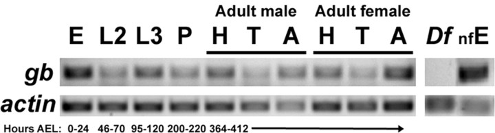
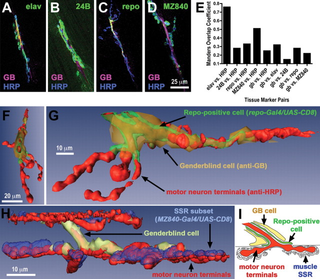
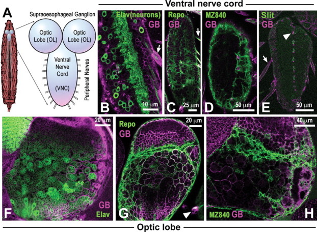
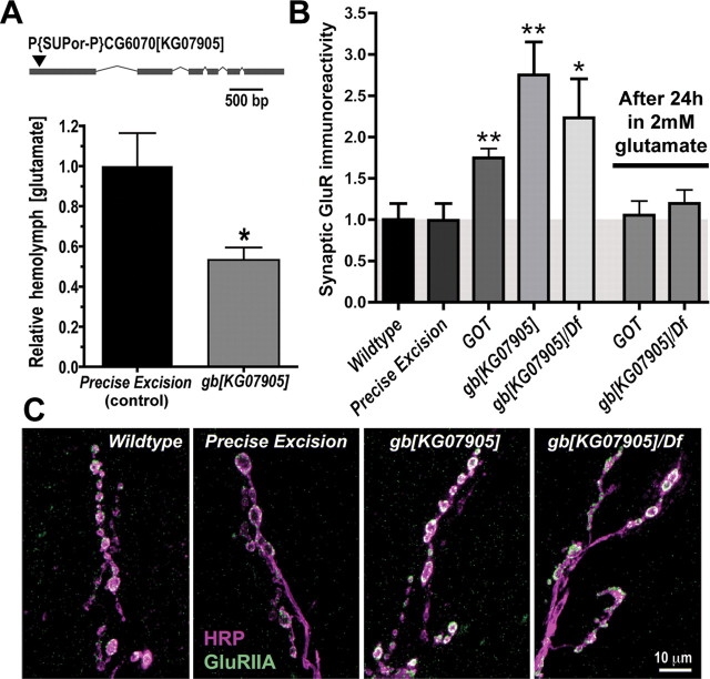
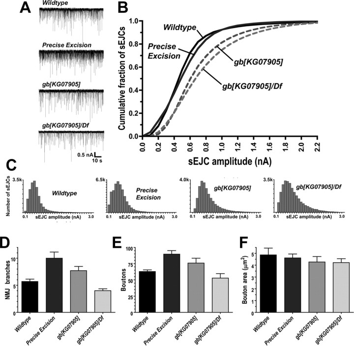
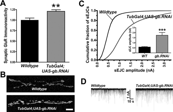
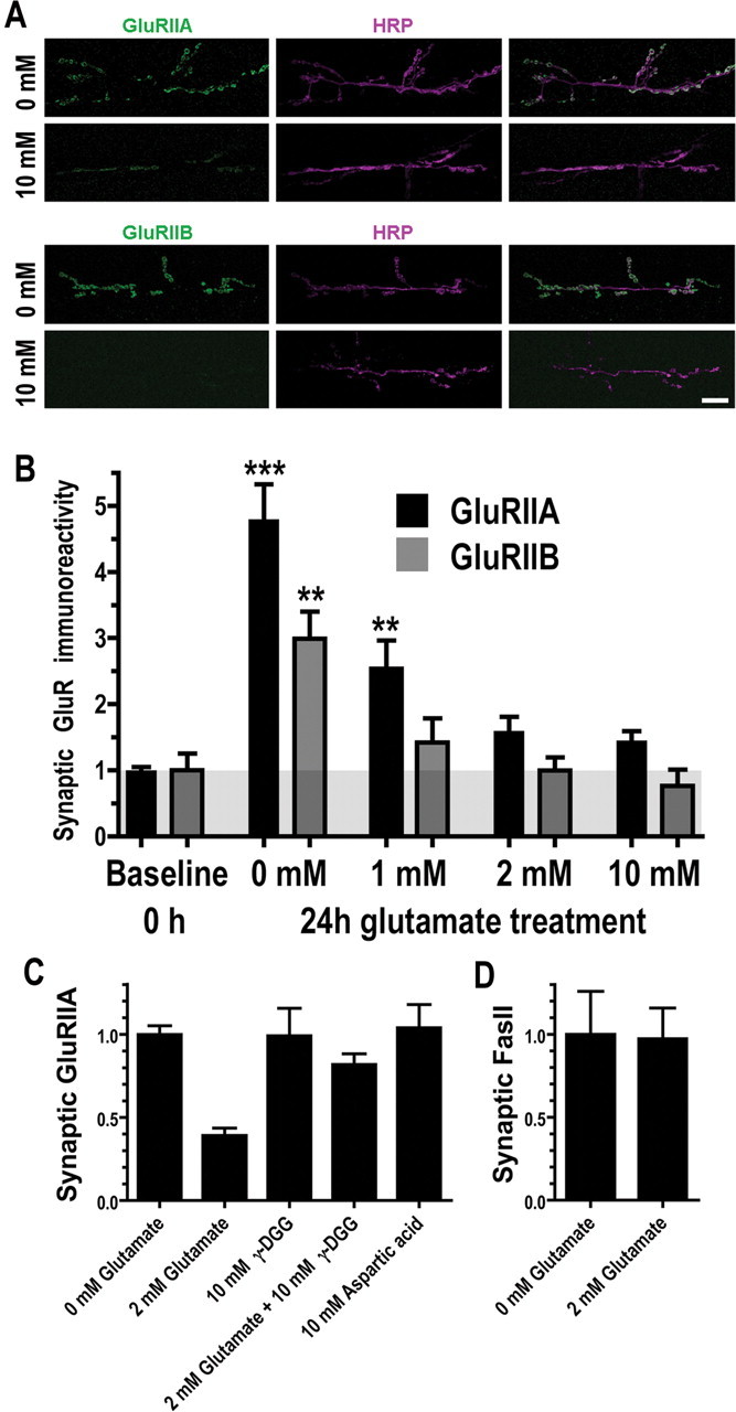
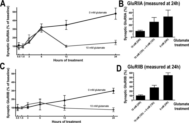
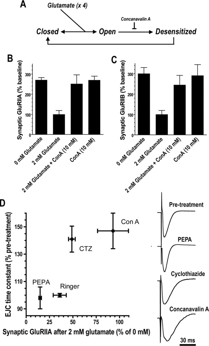
We hypothesized that cystine/glutamate transporters (xCTs) might be critical regulators of ambient extracellular glutamate levels in the nervous system and that misregulation of this glutamate pool might have important neurophysiological and/or behavioral consequences. To test this idea, we identified and functionally characterized a novel Drosophila xCT gene, which we subsequently named "genderblind" (gb). Genderblind is expressed in a previously overlooked subset of peripheral and central glia. Genetic elimination of gb causes a 50% reduction in extracellular glutamate concentration, demonstrating that xCT transporters are important regulators of extracellular glutamate. Consistent with previous studies showing that extracellular glutamate regulates postsynaptic glutamate receptor clustering, gb mutants show a large (200-300%) increase in the number of postsynaptic glutamate receptors. This increase in postsynaptic receptor abundance is not accompanied by other obvious synaptic changes and is completely rescued when synapses are cultured in wild-type levels of glutamate. Additional in situ pharmacology suggests that glutamate-mediated suppression of glutamate receptor clustering depends on receptor desensitization. Together, our results suggest that (1) xCT transporters are critical for regulation of ambient extracellular glutamate in vivo; (2) ambient extracellular glutamate maintains some receptors constitutively desensitized in vivo; and (3) constitutive desensitization of ionotropic glutamate receptors suppresses their ability to cluster at synapses.


Figures

Figure 1.
Figure 1.
GB is expressed throughout development. Representative RT-PCR measurements of relative genderblind and actin 5C transcript levels throughout development and in specific regions of adult male and female bodies. An equal quantity of RNA (measured spectrophotometrically) was isolated from each designated tissue type and used to produce the RT-PCR product shown in each lane. E, Embryos, L2, second-instar larvae; P, pupae; H, adult head; T, adult thorax; A, adult abdomen; Df, first-instar larval Df(3R)Exel6206 mutants (negative control); nfE, unfertilized embryos (showing heavy maternal gb RNA contribution). The age in hours after egg laying (AEL) at 25°C is shown below the lane images.
Figure 2.
Figure 2.
GB is expressed in a specific subset of NMJ-associated glia. A–D, Confocal projections of third-instar larval NMJs on ventral longitudinal muscles 6 and 7, stained with antibodies against GB (magenta), HRP (which allows visualization of all neuronal membrane; blue), and CD8 (green). For each image, transgenic transmembrane protein CD8::GFP was expressed in a specific tissue type using the gal4/UAS system. A, CD8 expression was driven with the pan-neuronal driver elav–Gal4 (elav). B, CD8 was driven with the muscle driver 24B–Gal4 (24B). C, CD8 was driven with the glial subset driver repo–Gal4 (repo). D, CD8 was driven with the weak larval muscle and CNS glial driver MZ840–Gal4 (MZ840). E, Quantitation of overlap between the different markers of specific tissue membranes described above. Elav versus HRP is a positive control, because elav–Gal4;UAS–CD8::GFP stained with anti-CD8 and anti-HRP antibodies both mark neuronal membranes. 24B versus HRP is a negative control, because 24B–Gal4;UAS–CD8::GFP stained with anti-CD8 antibodies stains muscle membrane, and anti-HRP stains neuronal membrane. F, 3D isosurface reconstruction of a 6/7 NMJ, stained as in C and viewed from the same perspective as the NMJs depicted in A–D. G, The same reconstruction shown in F, rotated, enlarged, and labeled. H, 3D isosurface reconstruction of a 6/7 NMJ stained as in D. I, Cross-sectional diagram of a larval NMJ, showing the arrangement of presynaptic motor neuron terminals, postsynaptic muscle membrane, and NMJ-associated glia.
Figure 3.
Figure 3.
GB is expressed in a specific subset of CNS glia. A, Diagram of neuromuscular anatomy in a third-instar larva. B–H, Confocal projections of larval ventral ganglia, stained with antibodies against GB (magenta) and CD8 (green). B, CD8 expression was driven with the pan-neuronal driver elav–Gal4 (Elav). C, CD8 was driven with the glial subset driver repo–Gal4 (Repo). D, CD8 was driven with MZ840–Gal4 (MZ840). E, The VNC is stained with antibodies against GB (magenta) and the midline glia marker slit (green). Arrows in B, C, and E indicate peripheral glial ensheating segmental nerves. Arrowhead in E indicates a midline glial cell expressing GB. F–H, Confocal projections of larval optic lobes, stained with antibodies against GB (magenta) and CD8 (green). CD8 was driven under control of elav–Gal4 (F), repo–Gal4 (G), or MZ840–Gal4 (H), as labeled and described above. Arrowhead in G indicates GB expression in an unidentified structure near the optic lobes.
Figure 4.
Figure 4.
genderblind mutants show an increase in postsynaptic glutamate receptor immunoreactivity. A, Relative concentration of glutamate in larval hemolymph, in gb mutants and precise-excision controls. gb[KG07905] mutants represent animals homozygous for a transposon insertion in the first exon of the gb gene (A, top). Precise-excision animals represent animals homozygous for the same chromosome after precise excision of the transposon (confirmed by sequencing of gb). n for each genotype equals 10 independently analyzed samples derived from 50 larvae. To control for variability in hemolymph extraction and/or evaporation, we normalized glutamate concentration measured in each sample by dividing it by the concentration of free phenylalanine in the same sample (see Materials and Methods). B, gb mutants show large increases in relative synaptic GluRIIA immunofluorescence, compared with controls (n = 6 per genotype). This large increase in GluR immunoreactivity is rescued when semi-intact gb and GOT mutants are cultured for 24 h in a physiologically normal (2 mm) hemolymph glutamate concentration. C, Portions of representative confocal micrographs showing larval NMJs on ventral longitudinal muscles 6/7, stained with anti-GluRIIA antibodies (green) and the neuronal membrane marker anti-HRP (magenta). White indicates overlap. Error bars represent SEM.
Figure 5.
Figure 5.
genderblind mutants show an increase in the number of functional postsynaptic glutamate receptors. A, Portions of two-electrode voltage-clamp recordings from the L3 muscle 6 NMJ in various genotypes, showing sEJCs (downward deflections). B, Cumulative frequency histogram of sEJC amplitudes from various genotypes; a right shift of the curve represents larger synaptic currents (n = 10–13 animals, ∼18,000–41,000 events measured per genotype). C, Frequency histograms of sEJC amplitudes from various genotypes. k, 1000. D–F, Number of presynaptic branches (D) and presynaptic boutons (E) and area of individual boutons (F) at the 6/7 NMJ, for various genotypes (n = 6–8 animals per genotype). Error bars represent SEM.
Figure 6.
Figure 6.
Transgenic expression of genderblind RNAi phenocopies gb mutants. A, Relative synaptic GluRIIA immunoreactivity (as in Fig. 4B) after knockdown of gb by ubiquitous expression of gb RNAi. B, Representative GluRIIA staining (as in Fig. 4C). Scale bar, 10 μm. C, Cumulative frequency histograms of sEJC amplitudes (as in Fig. 5B). D, Representative electrophysiological recordings (as in Fig. 5A; n = 10–13 animals). Error bars represent SEM.
Figure 7.
Figure 7.
Glutamate negatively regulates postsynaptic glutamate receptor abundance. A, Confocal micrographs of WT 6/7 NMJs cultured for 24 h in culture medium containing 0 or 10 mm glutamate and then fixed and stained with antibodies against glutamate receptor subunits GluRIIA (top, green) or GluRIIB (bottom, green) and the neuronal membrane marker anti-HRP (magenta). Scale bar, 10 μm. B, Postsynaptic 6/7 NMJ glutamate receptor immunofluorescence in larvae cultured in medium containing various amounts of glutamate. A large increase in postsynaptic glutamate receptors was measured when larvae were cultured for 24 h in medium containing <2 mm glutamate (n = 8–16 animals per genotype per assay). C, Glutamate-dependent loss of postsynaptic glutamate receptors is blocked by 10 mm γ-DGG, a competitive glutamate receptor antagonist, and does not occur in response to 10 mm aspartic acid (n = 5–23 animals per genotype per assay). D, Glutamate does not trigger loss of the presynaptic and postsynaptic transmembrane protein fasciclin II (FasII; n = 6–8 animals). Error bars represent SEM.
Figure 8.
Figure 8.
Time course of glutamate-dependent changes in postsynaptic glutamate receptor abundance. A, Synaptic GluRIIA immunoreactivity after a semi-intact neuromuscular preparation is cultured in glutamate for varying amounts of time and then immediately fixed and stained. B, Changes in GluRIIA immunoreactivity after 24 h when preparations are kept in 10 mm glutamate for the entire 24 h (first bar), kept in 10 mm glutamate for 12 h and then 0 mm glutamate for the next 12 h (second bar), or kept in 0 mm glutamate for the entire 24 h (third bar). C, Glutamate-dependent changes in postsynaptic GluRIIB immunoreactivity, as in A. D, Changes in GluRIIB immunoreactivity, as in B (n = 5–21 animals per time point). Error bars represent SEM.
Figure 9.
Figure 9.
Glutamate suppresses glutamate receptor localization via a desensitization-dependent mechanism. A, Diagrammatic summary of ionotropic glutamate receptor gating: binding of glutamate to each subunit causes receptor pore opening, followed by desensitization. B, Glutamate-dependent loss of A-type postsynaptic glutamate receptors is blocked by 10 mm concanavalin A (ConA), a desensitization inhibitor (n = 5–20 animals). C, Glutamate-dependent loss of B-type postsynaptic glutamate receptors is blocked by 10 mm concanavalin A (n = 5–20 animals). D, Different pharmacological agents block glutamate-dependent loss of synaptic GluRIIA in inverse proportion to the degree with which the agents inhibit desensitization (n = 4–20 animals). The ability of each agent to inhibit desensitization was quantified by measuring the increase in EJC falling phase time constants caused by drug application. Sample EJC recordings are shown on the right.

Similar articles

Cited by

References

    1. Aldred S, Moore KM, Fitzgerald M, Waring RH. Plasma amino acid levels in children with autism and their families. J Autism Dev Disord. 2003;33:93–97. - PubMed
    1. Armstrong N, Jasti J, Beich-Frandsen M, Gouaux E. Measurement of conformational changes accompanying desensitization in an ionotropic glutamate receptor. Cell. 2006;127:85–97. - PubMed
    1. Baker DA, Xi ZX, Shen H, Swanson CJ, Kalivas PW. The origin and neuronal function of in vivo nonsynaptic glutamate. J Neurosci. 2002;22:9134–9141. - PMC - PubMed
    1. Ball R, Xing B, Bonner P, Shearer J, Cooper RL. Long-term in vitro maintenance of neuromuscular junction activity of Drosophila larvae. Comp Biochem Physiol A Mol Integr Physiol. 2003;134:247–255. - PubMed
    1. Banerjee S, Pillai AM, Paik R, Li J, Bhat MA. Axonal ensheathment and septate junction formation in the peripheral nervous system of Drosophila. J Neurosci. 2006;26:3319–3329. - PMC - PubMed

Publication types

MeSH terms

LinkOut - more resources