. 2007 Jan 3;27(1):180-9.
doi: 10.1523/JNEUROSCI.3227-06.2007.

Critical period window for spectral tuning defined in the primary auditory cortex (A1) in the rat

Affiliations

Critical period window for spectral tuning defined in the primary auditory cortex (A1) in the rat

Etienne de Villers-Sidani et al. J Neurosci. .

Abstract

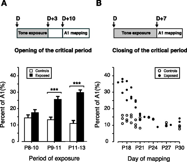
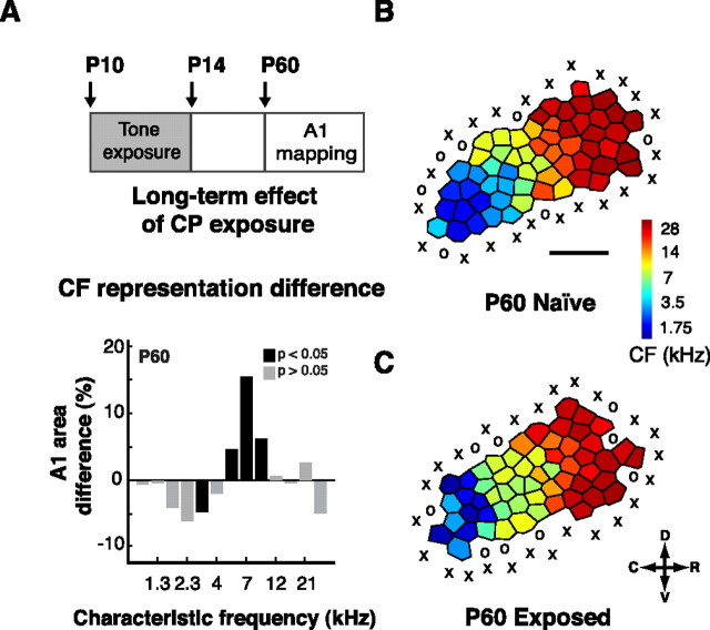
Experience-dependent plasticity during development results in the emergence of highly adapted representations of the external world in the adult brain. Previous studies have convincingly shown that the primary auditory cortex (A1) of the rat possesses a postnatal period of sensory input-driven plasticity but its precise timing (onset, duration, end) has not been defined. In the present study, we examined the effects of pure-tone exposure on the auditory cortex of developing rat pups at different postnatal ages with a high temporal resolution. We found that pure-tone exposure resulted in profound, persistent alterations in sound representations in A1 only if the exposure occurred during a brief period extending from postnatal day 11 (P11) to P13. We also found that postnatal sound exposure in this epoch led to striking alterations in the cortical representation of sound intensity.


Figures

Figure 1.
Figure 1.
Opening and closing of the CP. A, Top, Experimental protocol used to determine the time of opening of the CP. Rats were exposed to 3 d of 7 kHz pure tones starting at different postnatal days (D) and mapped 7 d after the end of the exposure period. Bottom, Percentage of A1 area with receptive fields tuned to 7 kHz ± 0.3 octave as a function of the exposure period (P8–P10 period, n = 5; P9–P11 period, n = 5; P11–P13 period, n = 5; naive, n = 4). Values shown are mean ± SE. ***p < 0.001, t test. B, Top, Experimental protocol used to determine the time of closing of the CP. Rats were exposed to 7 d of 7 kHz pure tones starting at different postnatal days (D) and mapped 7 d later (D+7). Bottom, Percentage of A1 area with receptive fields tuned to 7 kHz ± 0.3 octave, with respect to the day of mapping (D+7) (exposed, filled circles, n = 25; naive, open circles, n = 21).
Figure 2.
Figure 2.
Effect of CP exposure on A1 characteristic-frequency maps. A, B, Representative A1 CF map from a naive P18 rat (A) and from a rat exposed to 7 kHz pure tones throughout the critical period, then mapped at P18 (B). C, D, Distribution of tuning curve CFs plotted against a normalized tonotopic axis (see Materials and Methods) in litters of naive controls (C) and litters exposed during the CP (D). Note the increased number of recording sites at which neurons were tuned to 7 kHz in the exposed group (arrow). Scale bar, 0.5 mm. X, Unresponsive cortical site; O, non-AI cortical site (see Materials and Methods); D, dorsal; C, caudal; R, rostral; V, ventral. (exposed, n = 9; recorded sites, 458; naive, n = 9; recorded sites, 479.)
Figure 3.
Figure 3.
Long-term effect of CP exposure on A1 tuning. A, Top, Experimental protocol used to evaluate the long-term impact of CP exposure on A1 tuning. Rats were exposed to 5 d of 7 kHz pure tones starting 1 d before the opening of CP (P10) and ending 1 d after the closure (P14) to finally be mapped at P60. Bottom, Difference in frequency tuning between exposed and naive rats expressed as A1 percentage and separated by CF for litters mapped at P60 (controls, n = 4; exposed, n = 4). B, C, Representative A1 CF maps from a naive P60 rat (B) and from a rat exposed to 7 kHz pure tones throughout the critical period, between P10 and P14, and then mapped at P60 (C). Values shown are mean ± SE. **p < 0.01, t test. D, Dorsal; C, caudal; R, rostral; V, ventral.
Figure 4.
Figure 4.
Effect of CP exposure on frequency representation. A–C, Difference in frequency tuning between exposed and naive rats expressed as A1 percentage and separated by CF for litters mapped at P17 (A), P20 (B), and P23 (C). These rats were exposed for 7 d of 7 kHz pure tones and mapped on the seventh day of exposure (exposed, n = 9; naive, n = 9). D, E, Percentage of A1 activated by every frequency-intensity combination used for mapping in naive (D) and CP tone-exposed (E) rats. F, Difference in percent of activation between CP tone-exposed and naive rats (exposed, n = 9; naive, n = 9). **p < 0.01, t test.
Figure 5.
Figure 5.
Maturation of excitatory tone-evoked responses in A1 during the CP. A–D, Representative A1 CF maps obtained at different postnatal ages in naive controls between P11 and P14. Scale bar, 0.5 mm. X, Unresponsive cortical site; O, non-AI cortical site; D, dorsal; C, caudal; R, rostral; V, ventral. E–G, I–VI, Sites where the receptive fields were recorded. H–K, Repertoire of tuning curves in P11 (H), P12 (I), P13 (J), and P14 (K) naive rats. The apex of each pair of joined lines indicates the threshold and CF recorded at each particular penetration site. The separation 20 dB above that apex is the BW20 described in Materials and Methods and Results. (n = 3 for each postnatal day between P10 and P14.)
Figure 6.
Figure 6.
Evolution of response characteristics in A1 as a function of age. A, Bandwidths at 20 dB above threshold. B, Thresholds. C, Latencies. D, Total A1 cortical area. Open symbols, Cortical sites with a CF between 1–5.6 kHz; closed symbols, cortical sites with a CF between 5.7–30 kHz; age, day of mapping; all data are from naive (nontone-exposed) rats (n = 21).
Figure 7.
Figure 7.
Effect of CP exposure on response characteristics in A1. A–C, Bandwidths at 20 dB above threshold (A), thresholds (B), and onset latencies (C) separated by characteristic frequency in rats exposed (dark bars) throughout the CP (n = 9) and in naive (light bars) rats (n = 9).
Figure 8.
Figure 8.
CP exposure increases nonmonotonicity. A, Proportion of recorded sites per animal with nonmonotonic rate-level functions (see Materials and Methods) in A1 in exposed (E) and naive controls (C) grouped by frequency tuning (left, all sites recorded; middle, sites tuned to 7kHz ± 0.3 octave; right, non-7-kHz-tuned sites; exposed, n = 9; naïve, n = 9). B, C, Representative rate-level functions of a nonmonotonic site (B) and monotonic site (C) recorded in a tone-exposed animal and a naive control, respectively. m, Slope of segment between inflection point (arrow) and end of data. **p < 0.01, t test.

Similar articles

Cited by

References

    1. Astl J, Popelar J, Kvasnak E, Syka J. Comparison of response properties of neurons in the inferior colliculus of guinea pigs under different anesthetics. Audiology. 1996;35:335–345. - PubMed
    1. Bao S, Chang EF, Heiser MA, Merzenich MM. Representation of complex sounds through early experience. Soc Neurosci Abstr. 2003a;29:182.16.
    1. Bao S, Chang EF, Davis JD, Gobeske KT, Merzenich MM. Progressive degradation and subsequent refinement of acoustic representations in the adult auditory cortex. J Neurosci. 2003b;23:10765–10775. - PMC - PubMed
    1. Bao S, Chang EF, Woods J, Merzenich MM. Temporal plasticity in the primary auditory cortex induced by operant perceptual learning. Nat Neurosci. 2004;7:974–981. - PubMed
    1. Bartoletti A, Medini P, Berardi N, Maffei L. Environmental enrichment prevents effects of dark-rearing in the rat visual cortex. Nat Neurosci. 2004;7:215–216. - PubMed

Publication types