. 2007 Jan 24;26(2):288-300.
doi: 10.1038/sj.emboj.7601493. Epub 2007 Jan 4.

Cex1p is a novel cytoplasmic component of the Saccharomyces cerevisiae nuclear tRNA export machinery

Affiliations

Cex1p is a novel cytoplasmic component of the Saccharomyces cerevisiae nuclear tRNA export machinery

Andrew T McGuire et al. EMBO J. .

Abstract

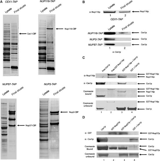
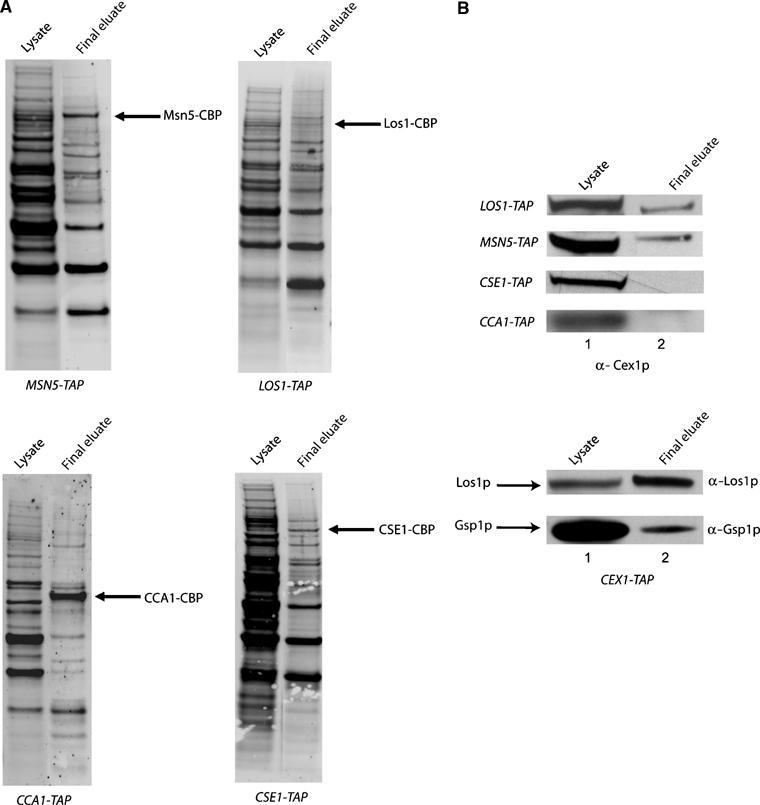
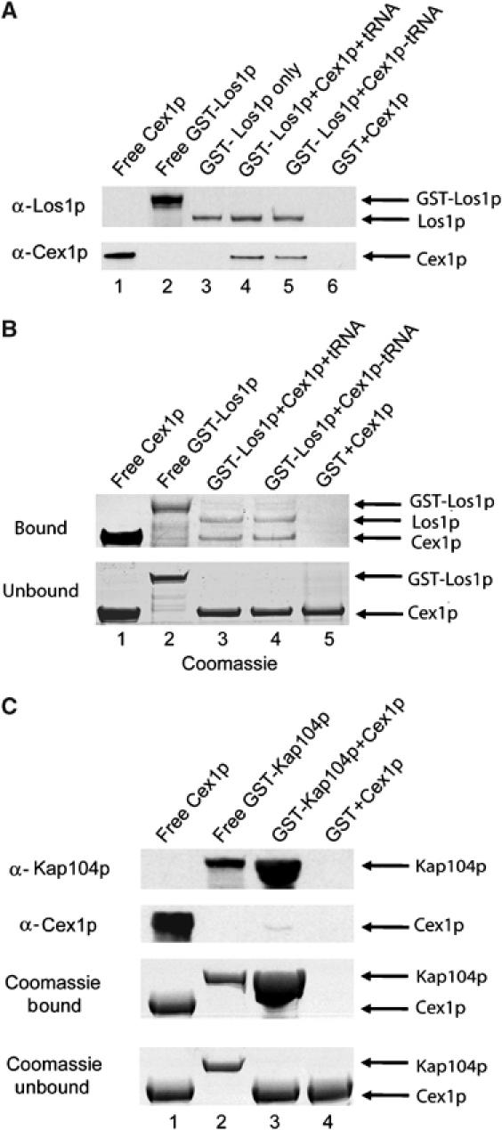
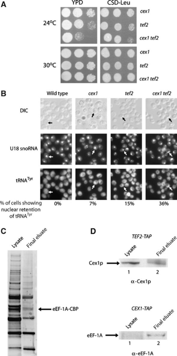
The Saccharomyces cerevisiae Yor112wp, which we named Cex1p, was identified using a yeast tRNA three-hybrid interaction approach and an in vivo nuclear tRNA export assay as a cytoplasmic component of the nuclear tRNA export machinery. Cex1p binds tRNA saturably, and associates with the nuclear pore complex by interacting directly with Nup116p. Cex1p co-purifies with the nuclear tRNA export receptors Los1p and Msn5p, the eukaryotic elongation factor eEF-1A, which delivers aminoacylated tRNAs to the ribosome, and the RanGTPase Gsp1p, but not with Cca1p, a tRNA maturation enzyme that facilitates translocation of non-aminoacylated tRNAs across the nuclear pore complex. Depletion of Cex1p and eEF-1A or Los1p significantly reduced the efficiency of nuclear tRNA export. Cex1p interacts with Los1p but not with eEF-1A in vitro. These findings suggest that Cex1p is a component of the nuclear aminoacylation-dependent tRNA export pathway in S. cerevisiae. They also suggest that Cex1p collects aminoacyl-tRNAs from the nuclear export receptors at the cytoplasmic side of the nuclear pore complex, and transfers them to eEF-1A using a channelling mechanism.


Figures

Figure 1
Figure 1
Disruption of the CEX1 and ARC1 genes reduced cell growth, which could be rescued by Arc1p but not by Los1p. The cex1 arc1, arc1 and cex1 strains containing pCEN-URA-GAL1-CEX1 were streaked on CSD-Ura medium and incubated at 30°C. The growth of the cex1 arc1 strain harboring the pCEN-URA-GAL1-CEX1 plasmid on CS medium containing galactose was also monitored (A). The cex1 arc1 pCEN-URA-GAL1-CEX1 strain containing pUN100 without or with the ARC1 gene, and the arc1 pCEN-URA-GAL1-CEX1 and cex1 pCEN-URA-GAL1-CEX1 strains containing pUN100 were streaked on CSD medium lacking Leu and containing 0.1% 5-FOA and incubated at 30°C (B). The cex1 arc1 pCEN-URA-GAL1-CEX1 strain containing pYX242 without or with the LOS1 gene was streaked on CSD medium lacking Leu and containing 0.1% 5-FOA and incubated at 30°C (C).
Figure 2
Figure 2
Disruption of the CEX1 and LOS1 genes did not affect cell growth. The cex1, los1 and cex1 los1 strains containing pYX242 were serially diluted and spotted on YPD or CSD medium lacking Leu and incubated at 24, 30 and 37°C.
Figure 3
Figure 3
Depletion of both Cex1p and Los1p significantly reduced the efficiency of nuclear tRNA export. The location of the U18 snoRNA and the mature form of tRNATyr in each strain was detected by FISH. Cells showing nuclear retention of tRNATyr, as determined by U18 snoRNA localization, are indicated by arrows. The number of cells showing nuclear retention of tRNATyr was expressed as a percentage of total cells. For each strain, a total of 200 cells were inspected for nuclear accumulation of tRNATyr.
Figure 4
Figure 4
Cex1p binds tRNA directly and saturably. Substrate-induced intrinsic fluorescence quenching of Trp residues was used to determine whether purified Cex1p (A) interacts with tRNA or DNA (B).
Figure 5
Figure 5
Cex1p associates with the NPC. The location of the Cex1-Myc fusion protein and Nup116p (A) or Nup2p (B) in cex1 harboring pRS416-ADH1-CEX1-MYC was determined by immunofluorescence confocal microscopy using a mouse monoclonal anti-Myc antibody and rabbit polyclonal anti-Nup2p and anti-Nup116p. Colocalization of Cex1p with Nup116p or Nup2p (arrow) was determined by overlay analysis of the images. The location of Cex1-Myc and Nup2p in nup120 (C) and nup116 (D) with pRS416-ADH1-CEX1-MYC was determined as above and overlay analyses of the images were performed to ascertain whether Cex1p colocalizes with Nup2p.
Figure 6
Figure 6
Cex1p associates with the NPC by interacting directly and specifically with Nup116p. Total cell extract was prepared from the CEX1-TAP, NUP116-TAP, NUP2-TAP or NUP57-TAP strain and subjected to TAP. The proteins in the total cell lysate (lane 1) and the final TAP eluate (lane 2) were on separated a 4–12% Novex gel and transferred to nitrocellulose membrane. The blot was stained with Sypro-Ruby to detect co-purifying proteins (A), or probed with anti-Cex1p or anti-Nup116p (B). To investigate whether Cex1p interacts with Nup116p directly, GST-Nup116p (200 μg, 1.72 nmol) (lanes 2 and 3) or GST was bound to glutathione resin (lane 4) (C) and incubated with a two-fold molar excess of Cex1p (291 μg, 3.44 nmol). After the resin was washed, Nup116p was released from GST using TEV. Coomassie blue staining of an SDS–PAGE gel (third row) or Western blot analysis was used to detect Nup116p (first row) and Cex1p (second row) in an aliquot of the eluate. Purified Cex1p (lane 1) and GST-Nup116p (lane 2) were used as size markers. The unbound Cex1p in an aliquot of the wash eluate was detected by Coomassie blue staining of an SDS–PAGE gel (fourth row). The specificity of the interaction between Cex1p and Nup116p was tested by investigating whether Cex1p interacts with Nup57p (D). The analysis was conducted as described above except the amount of GST-Nup57p (200 μg, 3.44 nmol) and Cex1p (590 μg, 6.88 nmol) was increased by two-fold. Bound GST-Nup57p was released by boiling the resin in the SDS–PAGE buffer, and an aliquot was subjected to SDS–PAGE. Coomassie blue staining (third row) or Western blot analysis was used to detect Nup57p (first row) and Cex1p (second row). Purified Cex1p (lane 1) and GST-Nup57p (lane 2) were used as size markers. Unbound Cex1p in an aliquot of the wash eluate was detected by Coomassie blue staining of an SDS–PAGE gel (fourth row).
Figure 7
Figure 7
Cex1p co-purifies with Los1p, Msn5p and Gsp1p but not with Cse1p or Cca1p by TAP. TAP was performed using total cell extract prepared from CEX1-TAP, LOS1-TAP, MSN5-TAP, CSE1-TAP, GSP1-TAP and CCA1-TAP. Total cell extract (lane 1) and the final eluate (lane2) were subjected to SDS–PAGE and the separated proteins were transferred to nitrocellulose membranes. The membranes were stained with Sypro-Ruby to detect the co-purifying proteins (A), or used for Western blot analysis (B) to detect Cex1p, Los1p or Gsp1p.
Figure 8
Figure 8
Cex1p interacts directly and specifically with Los1p in a tRNA-independent manner in vitro. GST-Los1p (200 μg, 1.57 nmol) was bound to glutathione resin with (lane 4) or without tRNA (lanes 3 and 5), and incubated with buffer with (lanes 4 and 5) or without (lane 3) a two-fold molar excess of Cex1p (266 μg, 3.14 nmol). Cex1p was also incubated with bound GST (lane 6). The resin was washed and Los1p was released from bound GST using TEV. Western blot analysis was used to detect Los1p (A, top row) and Cex1p (A, bottom row) in an aliquot of the eluate. Purified Cex1p (lane 1) and GST-Los1p (Lane 2) were used as size markers. Coomassie blue staining of an SDS–PAGE gel was also used to monitor the extent of protein binding to Los1p with (lane 3) or without tRNA (lane 4) and GST (lane 5) (B, top row). Unbound Cex1p in an aliquot of the wash eluate was detected by Coomassie blue staining of an SDS–PAGE gel (B, bottom row). The specificity of the interaction of Cex1p with Los1p was tested by investigating whether Cex1p interacts with Kap104p (C). The analysis was performed as above except 1.92 nmol (200 μg) of GST-Kap104p and 3.84 nmol (327 μg) of Cex1p were used. The resin was boiled in SDS–PAGE sample buffer to release GST-Kap104p. Coomassie blue staining of an SDS–PAGE gel (third row) or Western blot analysis was used to detect Kap104p (top row) and Cex1p (second row) in an aliquot of the eluate. Purified Cex1p (lane 1) and GST-Kap104p (lane 2) were used as size markers. Unbound Cex1p in an aliquot of the wash eluate was detected by Coomassie blue staining of an SDS–PAGE gel (bottom panel).
Figure 9
Figure 9
Disruption of the CEX1 and TEF2 genes affected cell growth and the efficiency of nuclear tRNA export and TAP resulted in the co-isolation of Cex1p and eEF-1A. The cex1, tef2 and cex1 tef2 strains containing pYX242 were serially diluted and spotted on YPD (left panels) or CSD medium lacking Leu (right panels) and incubated at 24 and 30°C (A). The location of the U18 snoRNA and the mature form of tRNATyr in the cex1, tef2 and cex1 tef2 strains was detected by FISH. Cells showing nuclear retention of tRNATyr are indicated by arrows as determined by U18 snoRNA localization. The number of cells showing nuclear retention of tRNATyr was expressed as a percentage of total cells. For each strain, a total of 200 cells were inspected for nuclear accumulation of tRNATyr (B). TAP was performed using total cell extract prepared from TEF2-TAP and CEX1-TAP. The proteins in total cell lysate (lane 1) and the final eluate (lane 2) were separated by SDS–PAGE and transferred to nitrocellulose membrane. The membrane was stained with Sypro-Ruby to detect the proteins (C) or used for Western blot analysis to detect Cex1p (D, top row) or eEF-1A (D, bottom row).

Similar articles

Cited by

References

    1. Aitchison JD, Blobel G, Rout MP (1995) Nup120p: a yeast nucleoporin required for NPC distribution and mRNA transport. J Cell Biol 131: 1659–1675 - PMC - PubMed
    1. Arts GJ, Fornerod M, Mattaj IW (1998a) Identification of a nuclear export receptor for tRNA. Curr Biol 8: 305–314 - PubMed
    1. Arts GJ, Kuersten S, Romby P, Ehresmann B, Mattaj IW (1998b) The role of exportin-t in selective nuclear export of mature tRNAs. EMBO J 17: 7430–7441 - PMC - PubMed
    1. Azad AK, Stanford DR, Sarkar S, Hopper AK (2001) Role of nuclear pools of aminoacyl-tRNA synthetases in tRNA nuclear export. Mol Biol Cell 12: 1381–1392 - PMC - PubMed
    1. Bohnsack MT, Regener K, Schwappach B, Saffrich R, Paraskeva E, Hartmann E, Gorlich D (2002) Exp5 exports eEF1A via tRNA from nuclei and synergizes with other transport pathways to confine translation to the cytoplasm. EMBO J 21: 6205–6215 - PMC - PubMed

Publication types

MeSH terms