Pedicle morphology of the thoracic spine in preadolescent idiopathic scoliosis: magnetic resonance supported analysis
- PMID: 17203274
- PMCID: PMC2200789
- DOI: 10.1007/s00586-006-0281-y
Pedicle morphology of the thoracic spine in preadolescent idiopathic scoliosis: magnetic resonance supported analysis
Abstract
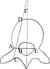
Although several studies have been reported on the adult vertebral pedicle morphology, little is known about immature thoracic pedicles in patients with idiopathic scoliosis. A total of 310 pedicles (155 vertebrae) from T1 to T12 in 10-14 years age group were analyzed with the use of magnetic resonance imaging and digital measurement program in 13 patients with right-sided thoracic idiopathic scoliosis. Each pedicle was measured in the axial and sagittal planes including transverse and sagittal pedicle width and angles, chord length, interpedicular distance and epidural space width on convex and concave sides of the curve. The smallest transverse pedicle widths were in the periapical region and the largest were in the caudal region. No statistically significant difference in transverse pedicle widths was detected between the convex and concave sides. The transverse pedicle angle measured 15.56 degrees at T1 and decreased to 6.32 degrees at T12. Chord length increased gradually from the cephalad part of the thoracic spine to the caudad part as the shortest length was seen at T1 convex level with a mean of 30.45 mm and the largest length was seen at T12 concave level with a mean of 41.73 mm. The width of epidural space on the concave side was significantly smaller than that on the convex side in most levels of the curve. Based on the anatomic measurements, it may be reasonable to consider thoracic pedicle screws in preadolescent idiopathic scoliosis.
Figures


References
-
- Dickson RA, Lawton JO, Archer IA, Butt WP. The pathogenesis of idiopathic scoliosis: biplanar spinal asymmetry. J Bone Joint Surg Br. 1987;66:8–15. - PubMed
MeSH terms
LinkOut - more resources
Full Text Sources
Medical

