Comparative Study
. 2007 Apr 1;55(5):453-62.
doi: 10.1002/glia.20467.

Systemic LPS causes chronic neuroinflammation and progressive neurodegeneration

Affiliations
Comparative Study

Systemic LPS causes chronic neuroinflammation and progressive neurodegeneration

Liya Qin et al. Glia. .

Abstract

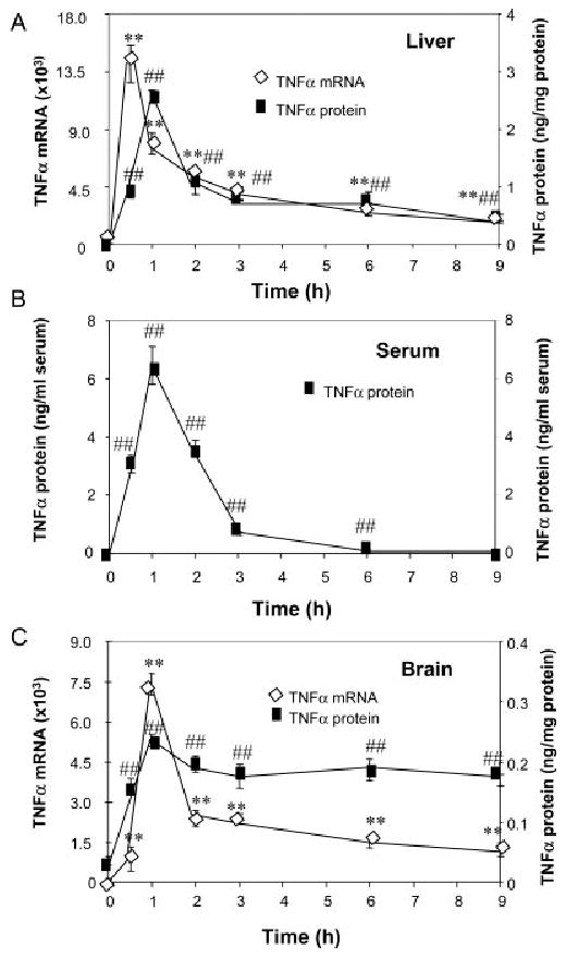
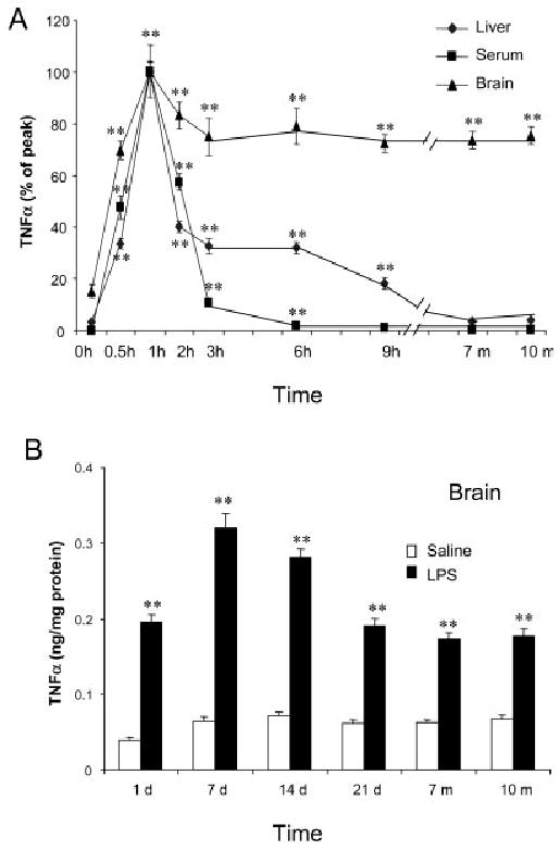
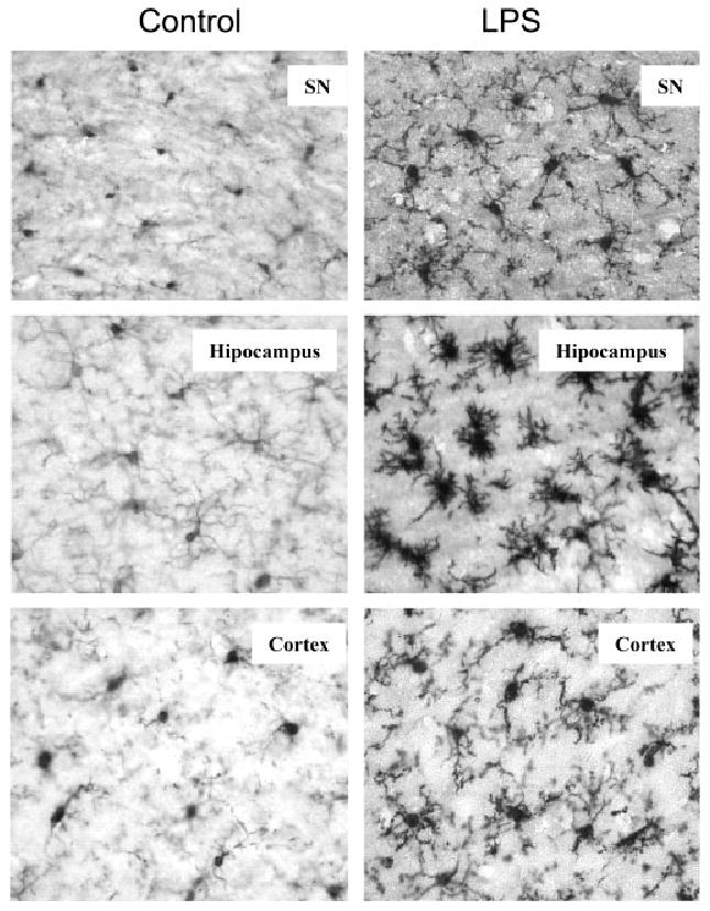
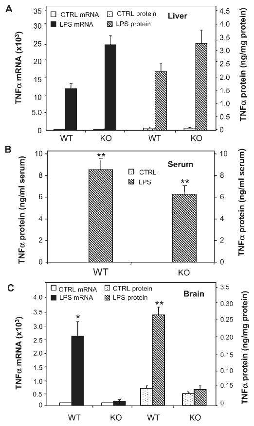
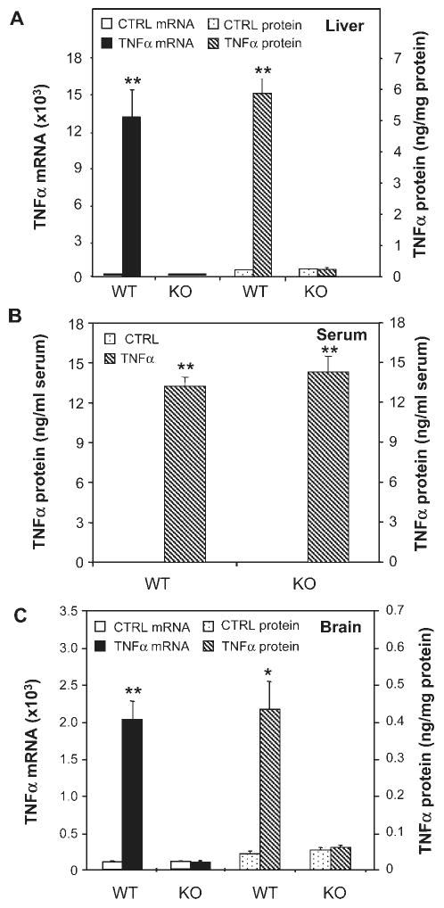
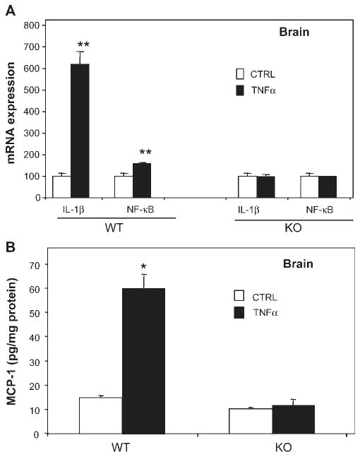
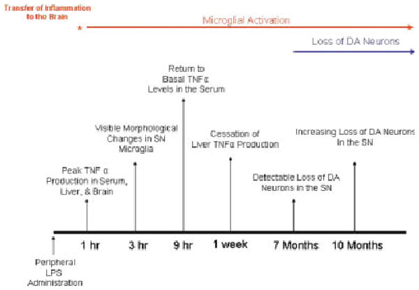
Inflammation is implicated in the progressive nature of neurodegenerative diseases, such as Parkinson's disease, but the mechanisms are poorly understood. A single systemic lipopolysaccharide (LPS, 5 mg/kg, i.p.) or tumor necrosis factor alpha (TNFalpha, 0.25 mg/kg, i.p.) injection was administered in adult wild-type mice and in mice lacking TNFalpha receptors (TNF R1/R2(-/-)) to discern the mechanisms of inflammation transfer from the periphery to the brain and the neurodegenerative consequences. Systemic LPS administration resulted in rapid brain TNFalpha increase that remained elevated for 10 months, while peripheral TNFalpha (serum and liver) had subsided by 9 h (serum) and 1 week (liver). Systemic TNFalpha and LPS administration activated microglia and increased expression of brain pro-inflammatory factors (i.e., TNFalpha, MCP-1, IL-1beta, and NF-kappaB p65) in wild-type mice, but not in TNF R1/R2(-/-) mice. Further, LPS reduced the number of tyrosine hydroxylase-immunoreactive neurons in the substantia nigra (SN) by 23% at 7-months post-treatment, which progressed to 47% at 10 months. Together, these data demonstrate that through TNFalpha, peripheral inflammation in adult animals can: (1) activate brain microglia to produce chronically elevated pro-inflammatory factors; (2) induce delayed and progressive loss of DA neurons in the SN. These findings provide valuable insight into the potential pathogenesis and self-propelling nature of Parkinson's disease.


Figures

Fig. 1
Fig. 1
Effects of LPS treatment on liver, plasma, and brain TNFα. Male C57BL/6 mice were treated with saline or LPS (5 mg/kg) i.p. injection. At different time points following LPS or saline injection, mice were sacrificed and brain extracts, liver extracts, and serum were prepared as described in methods. Analysis of TNFα mRNA and protein was conducted via real time RT-PCR and ELISA. (A) LPS rapidly increases liver TNFα mRNA and TNFα protein. (B) Serum TNFα protein is increased after LPS treatment. (C) Brain TNFα mRNA and TNFα protein are increased by LPS. The results are the means ± SEM of two experiments performed with five mice each time point. **P < 0.01, compared to the corresponding saline controls in TNFα mRNA data. ##P < 0.01, compared to the corresponding saline controls in TNFα protein data.
Fig. 2
Fig. 2
Single LPS injection caused long-lasting elevated level of TNFα protein in mouse brain. LPS (5 mg/kg) was injected intraperitoneally in male C57BL/6 mice. At different time points following LPS or saline injection, mice were sacrificed and brain extracts, liver extracts, and serum were prepared as described in methods. (A) Comparison of time course changes in TNFα protein in liver, serum and brain. Each tissue is adjusted to set peak values as 100%. Note that the liver and serum peak and decline in parallel, whereas, the brain remains elevated for at least 10 months. (B) Long term time course of LPS induced increases in TNFα protein in brain. TNFα levels peaked at 1 h and remained elevated for 10 months. The results are the means ± SEM of two experiments performed with five mice each time point.**P < 0.01, compared to the corresponding saline controls.
Fig. 3
Fig. 3
Immunocytochemical analysis of microglia. C57BL/6 mice were sacrificed 3 h following saline or LPS (5 mg/kg) i.p. injection. Brain sections were immnostained with Iba1 specific microglial antibody. Activated microglia in substantia nigra, hippocampus and cortex were shown by increased cell size, irregular shape, and intensified Iba1 staining in LPS-treated mouse brains. The images are from one experiment that is representative of three separate experiments.
Fig. 4
Fig. 4
Comparison of LPS-induced changes in TNFα in TNFR1/R2+/+ (WT) and TNFR1/R2−/− (KO) mice. TNFα mRNA and protein were measured 1 h after saline or LPS (5 mg/kg) i.p. injection. (A) LPS activated liver TNFα mRNA (left legend-solid bars) and TNFα protein (right legend-striped bars) in both wild type (WT) mice and mice lacking TNFα receptors (KO). (B) Serum TNFα protein is elevated by LPS in both WT and KO mice. (C) TNFα mRNA and protein was elevated in WT mice, but not in KO mice. These results indicate that TNFα receptors in brain are essential for LPS induced elevations of brain TNFα, but not for LPS induced elevations of liver TNFα. The results are the means ± SEM of two independent experiments performed with six mice for each treatment. *P < 0.05, **P < 0.01, compared to the corresponding saline controls.
Fig. 5
Fig. 5
Effects of systemic TNFα on liver, serum and brain TNFα in TNFR1/R2+/+ (WT) and TNFR1/R2−/− (KO) mice. Animals were sacrificed 1 h following saline or TNFα (0.25 mg/kg) i.p. injection. TNFα mRNA and protein was measured via real time RT-PCR and ELISA. (A) Wild type mice show a marked increase in liver TNFα mRNA (solid bars-left legend) and protein (striped bars-right legend) 1 h after TNFα treatment, but not TNFR1/R2 KO mice. (B) All mice injected with TNFα show increased serum levels of TNFα. (C) In wild type mice systemic TNFα injection markedly increases brain TNFα mRNA (solid bars-left legend) and protein (striped bars-right legend), but not TNFR1/R2 knock out mice. The results are the means ± SEM of two independent experiments performed with six mice for each treatment. *P < 0.05, **P < 0.01, compared to the corresponding saline controls.
Fig. 6
Fig. 6
Systemic TNFα increases IL-1β, NF-κB, and MCP-1 expression in TNFR1/R2+/+ (WT) mice. TNFR1/R2+/+ (WT) and TNFR1/R2−/− (KO) mice were injected with saline or TNFα (0.25 mg/kg) intraperitoneally. Brain mRNA expression of IL-1β and NF-κB-p65 and MCP-1 protein content were measured using real-time PCR and ELISA 1 h following saline or TNFα treatment. (A) Brain mRNA expression of IL-1β and NF-κB-p65 is increased by systemic TNFα in wild type mice, but not in TNFα receptor double KO mice. (B) Systemic TNFα administration increases brain MCP-1 protein in WT mice, but not in TNFR-KO mice. The results are the means ± SEM of two independent experiments performed with six mice for each treatment. *P < 0.05, **P < 0.01, compared to the corresponding saline control mice.
Fig. 7
Fig. 7
Delayed and time-dependent loss of nigral dopaminergic neurons following LPS i.p. injection. Male C57BL/6 mice were treated with saline or LPS (5 mg/kg, i.p.) and then maintained under normal conditions for the indicated time points. Mice were sacrificed and brain sections (35 mm thick) were cut through the nigral complex. After immunostaining with TH antibody, the number of TH-IR neurons in the SN was counted as described in methods. Results are expressed as percentage of the corresponding saline controls. (A) Number of TH-IR neurons in the SN of control and LPS-treated mice at different time points. (B) Visualization of TH-IR neurons in the substantia nigra and ventral tegmental area of saline and LPS-treated animals at 10 months. (C) F4/80-Immunohistochemistry for microglia within the SN and VTA of saline and LPS treated animals at 10 months. Note the high density of F4/80-IR microglia in the substantia nigra associated with the loss of dopaminergic neurons in mice treated with LPS. **P < 0.01, compared to the saline controls (n = 8–10 per time point).
Fig. 8
Fig. 8
Systemic LPS causes chronic microglial activation and progressive dopaminergic neurotoxicity. This figure chronologically depicts the pro-inflammatory profiles in the periphery and the brain in response to LPS and the consequences for dopaminergic neuron survival. TNFα levels peaked in serum, liver, and the brain at 1 h, indicating that transfer of inflammation from the periphery to the brain was rapid. Changes in microglia morphology indicative of activation were present at 3 h. However, while peripheral inflammation (serum and liver TNFα production) had subsided by 9 h (serum) and 1 week (liver) after LPS treatment, brain TNFα and microglial activation remained elevated for up to 10 months. Interestingly, significant loss of dopaminergic neurons was first detected only at 7 months after treatment and increased in severity at 10 months after LPS exposure. These events document the development of inflammation in the substantia nigra in response to peripheral LPS administration and the consequent initiation of delayed and progressive dopaminergic neurotoxicity.

Similar articles

Cited by

References

    1. Berkenbosch F, van Oers J, del Rey A, Tilders F, Besedovsky H. Corticotropin-releasing factor-producing neurons in the rat activated by interleukin-1. Science. 1987;238:524–526. - PubMed
    1. Bernardini R, Kamilaris TC, Calogero AE, Johnson EO, Gomez MT, Gold PW, Chrousos GP. Interactions between tumor necrosis factor-a, hypothalamic corticotropin-releasing hormone, and adrenocorticotropin secretion in the rat. Endocrinology. 1990;126:2876–2881. - PubMed
    1. Block ML, Hong JS. Microglia and inflammation-mediated neurodegeneration: Multiple triggers with a common mechanism. Prog Neurobiol. 2005;76:77–98. - PubMed
    1. Block ML, Wu X, Pei Z, Li G, Wang T, Qin L, Wilson B, Yang J, Hong JS, Veronesi B. Nanometer size diesel exhaust particles are selectively toxic to dopaminergic neurons: The role of microglia, phagocytosis, and NADPH oxidase. FASEB J. 2004;18:1618–1620. - PubMed
    1. Carvey PM, Chang Q, Lipton JW, Ling Z. Prenatal exposure to the bacteriotoxin lipopolysaccharide leads to long-term losses of dopamine neurons in offspring: A potential, new model of Parkinson's disease. Front Biosci. 2003;8:S826–S837. - PubMed

Publication types

MeSH terms