

Review
doi: 10.1111/j.1365-2559.2006.02544.x.
Infective disorders of the gastrointestinal tract
Affiliations
- PMID: 17204021
- PMCID: PMC7165686
- DOI: 10.1111/j.1365-2559.2006.02544.x
Item in Clipboard
Review
Infective disorders of the gastrointestinal tract
Histopathology.
2007 Jan.
Abstract
Gastrointestinal infections are a major cause of morbidity and mortality worldwide. Infectious organisms are often recovered by microbiological methods, but surgical pathologists may play a very valuable role in diagnosis. This review will focus on infective disorders of the gastrointestinal tract with an emphasis on enterocolitides caused by food- and water-borne pathogens. Diagnostic histological features of selected enteric infections will be emphasized, including those that mimic other inflammatory conditions of the gut (such as ischaemia or idiopathic inflammatory bowel disease), along with available diagnostic methods that can aid in diagnosis.
Figures
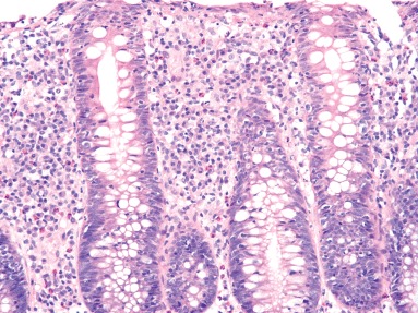
Numerous plasma cells in the lamina propria and increased intraepithelial lymphocytes may be seen in resolving infectious colitis, mimicking lymphocytic colitis (H&E).
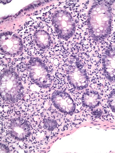
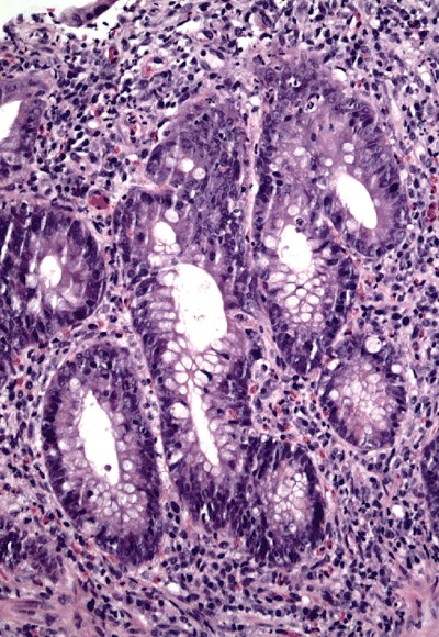
Culture and polymerase chain reaction‐proven Campylobacter colitis featuring well‐developed neutrophilic cryptitis, a neutrophilic infiltrate in the lamina propria and preservation of crypt architecture (H&E).
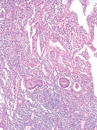
Salmonella typhi infection featuring mucosal ulceration, crypt abscesses and marked architectureal distortion, overlying a Peyer patch (H&E). Case courtesy of Dr A. Brian West.
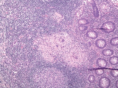
Epithelioid granulomas with prominent lymphoid cuffs in a case of polymerase chain reaction‐proven Yersinia enterocolitica (H&E).
Culture‐proven case of Aeromonas infection featuring focal cryptitis and architectural distortion from a right colon biopsy (H&E).
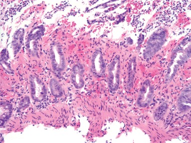
Biopsy from a case of Escherichia coli O157:H7 showing lamina propria hyalinization, crypt withering, neutrophilic inflammation in the mucosa and fibrin thrombi (H&E).
Similar articles
-
Pathology of food-borne infectious diseases of the gastrointestinal tract: an update.Adv Anat Pathol. 2003 Nov;10(6):319-27. doi: 10.1097/00125480-200311000-00002. Adv Anat Pathol. 2003. PMID: 14581821 Review.
-
Update on infectious enterocolitides and the diseases that they mimic.Histopathology. 2015 Jan;66(1):3-14. doi: 10.1111/his.12582. Histopathology. 2015. PMID: 25312041 Review.
-
Infectious Diseases of the Lower Gastrointestinal Tract.Surg Pathol Clin. 2010 Jun;3(2):297-326. doi: 10.1016/j.path.2010.05.009. Epub 2010 Aug 16. Surg Pathol Clin. 2010. PMID: 26839133 Review.
-
Pathology of inflammatory bowel disease.Semin Pediatr Surg. 2007 Aug;16(3):154-63. doi: 10.1053/j.sempedsurg.2007.04.005. Semin Pediatr Surg. 2007. PMID: 17602970 Review.
-
Gastrointestinal tract pathology in patients with common variable immunodeficiency (CVID): a clinicopathologic study and review.Am J Surg Pathol. 2007 Dec;31(12):1800-12. doi: 10.1097/PAS.0b013e3180cab60c. Am J Surg Pathol. 2007. PMID: 18043034
Cited by
-
Parvovirus b19 infection localized in the intestinal mucosa and associated with severe inflammatory bowel disease.J Clin Microbiol. 2009 May;47(5):1591-5. doi: 10.1128/JCM.00706-08. Epub 2009 Mar 11. J Clin Microbiol. 2009. PMID: 19279179 Free PMC article.
-
Immunohistochemistry and real-time Polymerase Chain Reaction: importance in the diagnosis of intestinal tuberculosis in a Peruvian population.BMC Gastroenterol. 2024 May 16;24(1):166. doi: 10.1186/s12876-024-03235-6. BMC Gastroenterol. 2024. PMID: 38755577 Free PMC article.
-
A Comprehensive Review of Infectious Granulomatous Diseases of the Gastrointestinal Tract.Gastroenterol Res Pract. 2021 Feb 6;2021:8167149. doi: 10.1155/2021/8167149. eCollection 2021. Gastroenterol Res Pract. 2021. PMID: 33628227 Free PMC article. Review.
-
Towards a new paradigm of microscopic colitis: Incomplete and variant forms.World J Gastroenterol. 2016 Oct 14;22(38):8459-8471. doi: 10.3748/wjg.v22.i38.8459. World J Gastroenterol. 2016. PMID: 27784958 Free PMC article. Review.
-
Human Mastadenovirus Infections in Children: A Review of the Current Status in the Arab World in the Middle East and North Africa.Children (Basel). 2022 Sep 6;9(9):1356. doi: 10.3390/children9091356. Children (Basel). 2022. PMID: 36138665 Free PMC article. Review.
References
-
- Surawicz CM. The role of rectal biopsy in infectious colitis. Am. J. Surg. Pathol. 1988; 12 (Suppl. 1); 82–88. - PubMed
-
- Abrams GD. Surgical pathology of the infected gut. Am. J. Surg. Pathol. 1987; 11 (Suppl. 1); 16–24. - PubMed
-
- Greenson JK, Stern RA, Carpenter SL, Barnett JL. The clinical significance of focal active colitis. Hum. Pathol. 1997; 28; 729–733. - PubMed
-
- Kumar NB, Nostrant TT, Appelman HD. The histopathologic spectrum of acute self‐limited colitis (acute infectious‐type colitis). Am. J. Surg. Pathol. 1982; 6; 523–529. - PubMed
-
- Fang G, Araujo V, Guerrant RL. Enteric infections associated with exposure to animals or animal products. Infect. Dis. Clin. N. Am. 1991; 5; 681–701. - PubMed
Publication types
MeSH terms
LinkOut - more resources
Full Text Sources
Medical
