

The neural circuitry underlying the executive control of auditory spatial attention
- PMID: 17204249
- PMCID: PMC3130498
- DOI: 10.1016/j.brainres.2006.11.088
The neural circuitry underlying the executive control of auditory spatial attention
Erratum in
- Brain Res. 2007 May 25;1147:284
Abstract
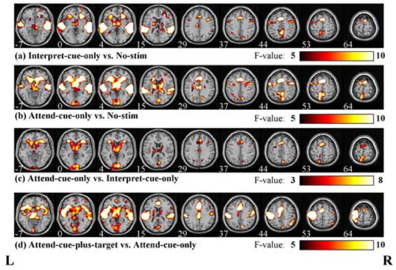
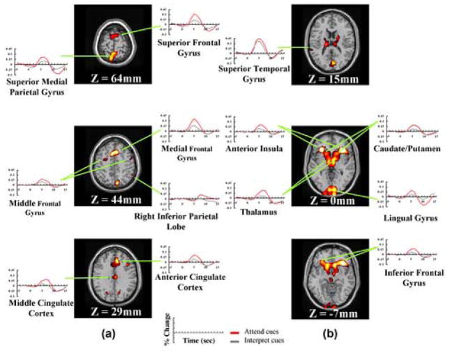
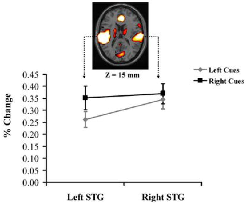
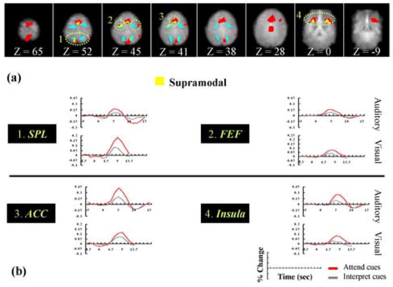
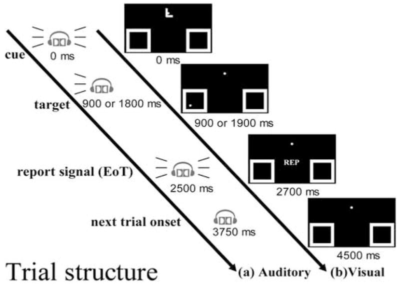
Although a fronto-parietal network has consistently been implicated in the control of visual spatial attention, the network that guides spatial attention in the auditory domain is not yet clearly understood. To investigate this issue, we measured brain activity using functional magnetic resonance imaging while participants performed a cued auditory spatial attention task. We found that cued orienting of auditory spatial attention activated a medial-superior distributed fronto-parietal network. In addition, we found cue-triggered increases of activity in the auditory sensory cortex prior to the occurrence of an auditory target, suggesting that auditory attentional control operates in part by biasing processing in sensory cortex in favor of expected target stimuli. Finally, an exploratory cross-study comparison further indicated several common frontal and parietal regions as being involved in the control of both visual and auditory spatial attention. Thus, the present findings not only reveal the network of brain areas underlying endogenous spatial orienting in the auditory modality, but also suggest that the control of spatial attention in different sensory modalities is enabled in part by some common, supramodal neural mechanisms.
Figures
Similar articles
-
Neuromagnetic recordings reveal the temporal dynamics of auditory spatial processing in the human cortex.Neurosci Lett. 2006 Mar 20;396(1):17-22. doi: 10.1016/j.neulet.2005.11.018. Epub 2005 Dec 15. Neurosci Lett. 2006. PMID: 16343772
-
Reorganisation of the right occipito-parietal stream for auditory spatial processing in early blind humans. A transcranial magnetic stimulation study.Brain Topogr. 2009 May;21(3-4):232-40. doi: 10.1007/s10548-009-0075-8. Epub 2009 Feb 6. Brain Topogr. 2009. PMID: 19199020
-
Fast and slow parietal pathways mediate spatial attention.Nat Neurosci. 2004 Mar;7(3):217-8. doi: 10.1038/nn1203. Epub 2004 Feb 22. Nat Neurosci. 2004. PMID: 14983182
-
Dissociating top-down attentional control from selective perception and action.Neuropsychologia. 2001;39(12):1277-91. doi: 10.1016/s0028-3932(01)00117-8. Neuropsychologia. 2001. PMID: 11566311 Review.
-
Representations of spectral coding in the human brain.Int Rev Neurobiol. 2005;70:331-69. doi: 10.1016/S0074-7742(05)70010-6. Int Rev Neurobiol. 2005. PMID: 16472639 Review. No abstract available.
Cited by
-
Toward a cumulative science of functional integration: A meta-analysis of psychophysiological interactions.Hum Brain Mapp. 2016 Aug;37(8):2904-17. doi: 10.1002/hbm.23216. Epub 2016 May 4. Hum Brain Mapp. 2016. PMID: 27145472 Free PMC article.
-
Dissociable influences of auditory object vs. spatial attention on visual system oscillatory activity.PLoS One. 2012;7(6):e38511. doi: 10.1371/journal.pone.0038511. Epub 2012 Jun 5. PLoS One. 2012. PMID: 22693642 Free PMC article. Clinical Trial.
-
fMRI evidence for both generalized and specialized components of attentional control.Brain Res. 2007 Oct 26;1177:90-102. doi: 10.1016/j.brainres.2007.07.097. Epub 2007 Sep 4. Brain Res. 2007. PMID: 17916338 Free PMC article.
-
Alignment of auditory artificial networks with massive individual fMRI brain data leads to generalisable improvements in brain encoding and downstream tasks.Imaging Neurosci (Camb). 2025 Apr 8;3:imag_a_00525. doi: 10.1162/imag_a_00525. eCollection 2025. Imaging Neurosci (Camb). 2025. PMID: 40800971 Free PMC article.
-
Neural pathways conveying novisual information to the visual cortex.Neural Plast. 2013;2013:864920. doi: 10.1155/2013/864920. Epub 2013 Jun 6. Neural Plast. 2013. PMID: 23840972 Free PMC article. Review.
References
-
- Arnell KM, Larson JM. Cross-modality attentional blinks without preparatory task-set switching. Psychon Bull Rev. 2002;9:497–506. - PubMed
-
- Behrmann M, Geng JJ, Shomstein S. Parietal cortex and attention. Curr Opin Neurobiol. 2004;14:212–217. - PubMed
-
- Bellmann A, Meuli R, Clarke S. Two types of auditory neglect. Brain. 2001;124:676–687. - PubMed
-
- Benedict RH, Shucard DW, Santa Maria MP, Shucard JL, Abara JP, Coad ML, Wack D, Sawusch J, Lockwood A. Covert auditory attention generates activation in the rostral/dorsal anterior cingulate cortex. J Cogn Neurosci. 2002;14:637–645. - PubMed
Publication types
MeSH terms
Grants and funding
LinkOut - more resources
Full Text Sources
