Comparative Study
. 2007 Mar 1;303(1):391-404.
doi: 10.1016/j.ydbio.2006.11.025. Epub 2006 Nov 21.

The role of TGF-beta signaling in regulating chondrogenesis and osteogenesis during mandibular development

Affiliations
Comparative Study

The role of TGF-beta signaling in regulating chondrogenesis and osteogenesis during mandibular development

Kyoko Oka et al. Dev Biol. .

Abstract

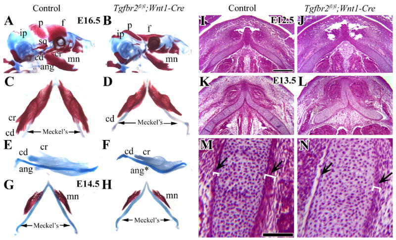
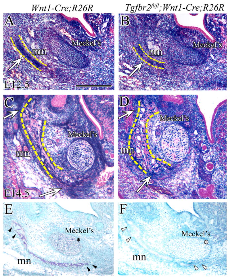
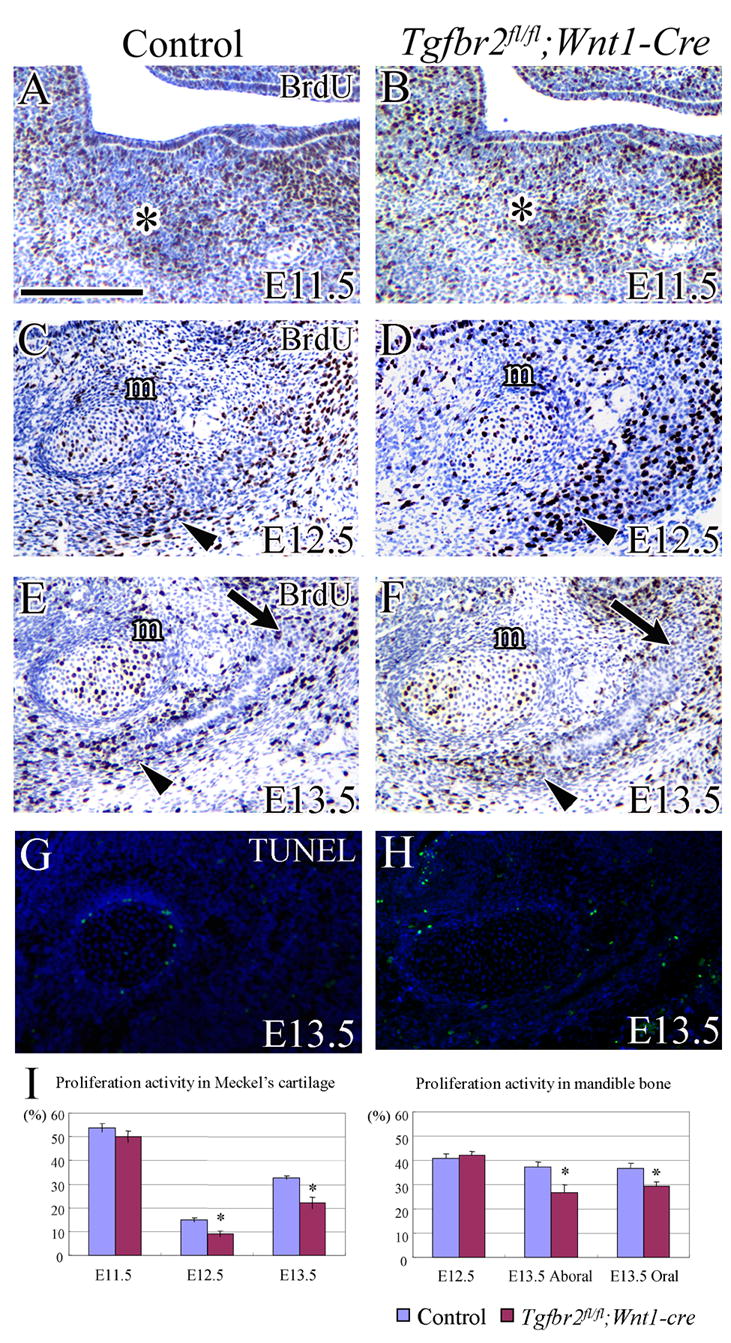
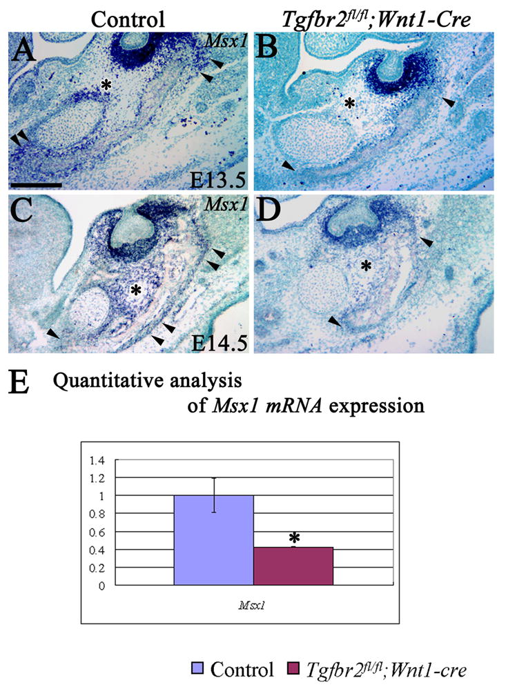
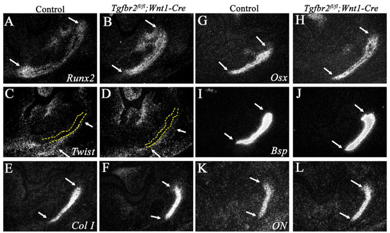
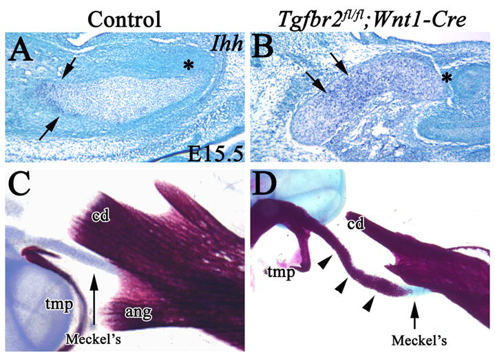
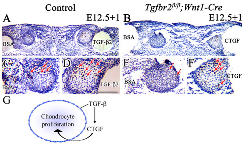
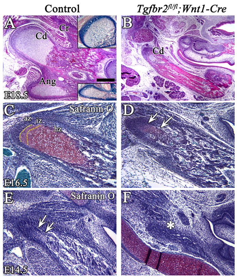
During craniofacial development, Meckel's cartilage and the mandible bone derive from the first branchial arch, and their development depends upon the contribution of cranial neural crest (CNC) cells. We previously demonstrated that conditional inactivation of Tgfbr2 in the neural crest of mice (Tgfbr2(fl/fl);Wnt1-Cre) results in severe defects in mandibular development, although the specific cellular and molecular mechanisms by which TGF-beta signaling regulates the fate of CNC cells during mandibular development remain unknown. We show here that loss of Tgfbr2 does not affect the migration of CNC cells during mandibular development. TGF-beta signaling is specifically required for cell proliferation in Meckel's cartilage and the mandibular anlagen and for the formation of the coronoid, condyle and angular processes. TGF-beta-mediated connective tissue growth factor (CTGF) signaling is critical for CNC cell proliferation. Exogenous CTGF rescues the cell proliferation defect in Meckel's cartilage of Tgfbr2(fl/fl);Wnt1-Cre mutants, demonstrating the biological significance of this signaling cascade in chondrogenesis during mandibular development. Furthermore, TGF-beta signaling controls Msx1 expression to regulate mandibular osteogenesis as Msx1 expression is significantly reduced in Tgfbr2(fl/fl);Wnt1-Cre mutants. Collectively, our data suggest that there are differential signal cascades in response to TGF-beta to control chondrogenesis and osteogenesis during mandibular development.


Figures

Figure 1
Figure 1. Developmental defects during the formation of Meckel’s cartilage and the mandible bone in Tgfbr2fl/fl;Wnt1-Cre mice
(A,B) Lateral view of skeletal staining preparations of control and Tgfbr2fl/fl;Wnt1-Cre mutant mice at E16.5. Tgfbr2fl/fl;Wnt1-Cre mice have severe defects of the frontal (f), parietal (p) and mandible bones (mn). (C,D) Top view of Meckel’s cartilage and the mandible bone at E16.5 in control and Tgfbr2fl/fl;Wnt1-Cre mutant mice. (E,F) Lateral view of mandible complexes stained with Alcian Blue at E16.5 in control and Tgfbr2fl/fl;Wnt1-Cre mutant mice. Tgfbr2fl/fl;Wnt1-Cre mice have a shortened mandible bone, diminished coronoid and condylar process, and absent angular process. (G,H) Top view of Meckel’s cartilage and the mandible bone at E14.5 in control and Tgfbr2fl/fl;Wnt1-Cre mutant mice samples. (I–M) Histological analysis of Meckel’s cartilage in transversal sections at E12.5 (I,J) and E13.5 (K–N). At E12.5, the Meckel’s cartilage in the Tgfbr2fl/fl;Wnt1-Cre mutant sample is indistinguishable from control. At E13.5, the Tgfbr2fl/fl;Wnt1-Cre mutant sample has defects including a curved Meckel’s cartilage and disrupted layers of the perichondrium and alignment of the chondrocytes in Meckel’s cartilage (arrows). The white parentheses indicate the thickness of the layers. Scale bars: 200μm in I–L; 100μm in M, N. f; frontal bone, p; parietal bone, ip; interparietal bone, sq; squamous bone, mn; mandible bone, cd; condylar process, cr; coronoid process, ang; angular process, Meckel’s; Meckel’s cartilage
Figure 2
Figure 2. CNC cell migration into Meckel’s cartilage is not affected by loss of Tgfbr2
CNC cells are (blue) visualized by X-gal staining of the mandible at E12.5 (A,B) and E14.5 (C,D) in R26R;Wnt1-Cre (control) and Tgfbr2fl/fl;R26R;Wnt1-Cre mice. (A,B) At E12.5, CNC-derived cells are detected in Meckel’s cartilage and the mandible primordium (arrow). There is no apparent difference in the population of cells in Meckel’s cartilage and mandible bone of R26R;Wnt1-Cre and Tgfbr2fl/fl;R26R;Wnt1-Cre mice. (C,D) At E14.5, the Meckel's cartilage is composed of CNC-derived and non-CNC-derived cells. The staining reveals the high density of CNC-derived cells in the osteogenic front of the mandible primordium (arrow). Yellow dashed lines indicate the mandible bone primordium (A,B) or the mandible bone (C,D). (E and F) In situ hybridization shows that Tgfbr2 is expressed (dark blue) in Meckel’s cartilage (*) and in the mandibular primordium (arrowhead) in the control (E) while it is not detectable in the Tgfbr2fl/fl;Wnt1-Cre sample (F) at E13.5. Scale bar: 200μm in A–D. mn; mandible, Meckel’s; Meckel’s cartilage.
Figure 3
Figure 3
Cell proliferation and apoptosis during Meckel’s cartilage and mandible bone development. (A–F) BrdU incorporation in the chondrocytes of Meckel’s cartilage and the osteogenic front of the mandible bone primordium in control and Tgfbr2fl/fl;Wnt1-Cre mice at E11.5, E12.5 and E13.5. BrdU staining (brown spot) indicates cell proliferation activity. (A,B) At E11.5, Meckel’s cartilage is visible as the highly condensed cell area (asterisk) in both control and Tgfbr2fl/fl;Wnt1-Cre mutant samples. (C,D) At E12.5, the Meckel’s cartilage (m) is well defined and the mandible bone primordium is becoming visible as the highly condensed cell area next to Meckel’s cartilage (arrowhead). (E,F) At E13.5, active CNC cell proliferation is seen in the oral (arrow) and aboral (arrowhead) side of the osteogenic front of the mandible bone, but it is significantly reduced in the Tgfbr2fl/fl;Wnt1-Cre mutant within both the oral and aboral areas. There is also a reduction of proliferation activity in Meckel’s cartilage (m). (G,H) TUNEL assay in the chondrocytes of Meckel’s cartilage and the mandible bone in control and Tgfbr2fl/fl;Wnt1-Cre mice at E13.5. Neither control nor Tgfbr2fl/fl;Wnt1-Cre mice have detectable apoptotic activity. (I) Quantification of cell proliferation activity from A–F. Asterisks indicate statistical significance (p<0.05). Scale bar: 200μm in A–H
Figure 4
Figure 4. Msx1 expression is down regulated in the Meckel’s cartilage and mandible bone of Tgfbr2fl/fl;Wnt1-Cre mice
(A–D) In situ hybridization of Msx1 mRNA within Meckel’s cartilage and the osteogenic front of the mandible bone (arrowhead) in control and Tgfbr2fl/fl;Wnt1-Cre mice at E13.5 and E14.5. (*) indicates Msx1 expression in A&C and no Msx1 expression in B&D. (E) Quantitative analysis of Msx1 mRNA expression by real time quantitative RT-PCR in Meckel’s cartilage at E13.5. Asterisks indicate statistical significance (p<0.01). Scale bar: 200μm in A–D
Figure 5
Figure 5. TGF-β signaling is not required for bone matrix maturation in the developing mandible bone
In situ hybridization of the mandible bone primordium at E13.5 in control and Tgfbr2fl/fl;Wnt1-Cre mice with Runx2 (A,B), Twist (C,D) Type I collagen (ColI; E,F), Osterix (Osx; G,H), Bsp (I,J), and Osteonectin (ON; K,L). Arrows indicate the oral (top) and aboral (bottom) sides of the osteogenic front of the mandible bone primordium. Yellow dashed lines (C,D) indicate the mandible bone.
Figure 6
Figure 6. Ctgf expression is down-regulated in the perichondrium of the Meckel’s cartilage in Tgfbr2fl/fl;Wnt1-Cre mice
(A–D) In situ hybridization of Ctgf in the perichondrium of Meckel’s cartilage at E12.5 and E13.5 in control and Tgfbr2fl/fl;Wnt1-Cre mice. There is a significant reduction of Ctgf expression in Tgfbr2fl/fl;Wnt1-Cre mice at E12.5 and E13.5 (arrowheads, A–D). (E) Quantitative analysis of Ctgf mRNA expression by real time quantitative RT-PCR in Meckel’s cartilage at E13.5. Ctgf expression is dramatically decreased in Tgfbr2fl/fl;Wnt1-Cre mutant samples. Asterisks indicate statistical significance (p<0.01). Scale bar: 200μm in A–D
Figure 7
Figure 7. Differentiation of chondrocytes in Meckel’s cartilage is accelerated in Tgfbr2fl/fl;Wnt1-Cre
(A,B) In situ hybridization of Ihh in Meckel’s cartilage of control and Tgfbr2fl/fl;Wnt1-Cre mice at E15.5. Ihh is expressed in the middle region (arrow) but not in the distal region (*) of Meckel’s cartilage in control mice. Ihh expression is expanded to the distal region of Meckel’s cartilage in Tgfbr2fl/fl;Wnt1-Cre mice. Asterisks indicate the distal end of Meckel’s cartilage. (C) Skeletal staining of the proximal part of the mandible in the control sample. (D) Skeletal staining of the proximal part of the mandible in Tgfbr2fl/fl;Wnt1-Cre mice. Abnormal ossification (arrowhead) is present around the proximal part of Meckel’s cartilage (arrow). Scale bar: 200μm in A–D. tmp, tympanic ring; cd, condylar process; Meckel’s, Meckel’s cartilage.
Figure 8
Figure 8. CTGF is a downstream mediator of TGF-β signaling to control cell proliferation in Meckel’s cartilage
BrdU labeling of cultured Meckel’s cartilage from control and Tgfbr2fl/fl;Wnt1-Cre mice at E12.5, treated with BSA, TGF-β2 or CFGF beads for 24 hours (E12.5+1). (A,C,D) Control sample treated with BSA and TGF-β2 beads. The number of BrdU labeled cells (red arrows) is much greater in Meckel’s cartilage treated with TGF-β2 beads. (B,E,F) Tgfbr2fl/fl;Wnt1-Cre sample treated with BSA and CTGF beads. The Tgfbr2fl/fl;Wnt1-Cre sample has few labeled cells with BSA beads but many more after treatment with CTGF beads (red arrows). (G) Schematic drawing demonstrates that TGF-β controls CTGF expression in the perichondrium to regulate chondrocyte proliferation during Meckel's cartilage development. Scale bar: 100μm in A–F.
Figure 9
Figure 9. TGF-β signaling is required for proper development of the condylar process
Histological analysis of the coronoid, condylar and angular processes in control and Tgfbr2fl/fl;Wnt1-Cre mice. (A,B) H&E staining shows clear zones of endochondral ossification in the condylar and angular processes in the control at E18.5. The insets show X-gal staining of the condyle and angular processes. The endochondral ossification of the condylar process is diminished in the Tgfbr2fl/fl;Wnt1-Cre mutant at E18.5. (C–F) Safranin O staining of the condyle process at E16.5 (C,D) and at E14.5 (E,F). At E16.5, the three zones, articular, intermediate, and hypertrophic, are visible in the control, but chondrogenesis of the condylar process is dramatically diminished and the hypertrophic zone is not detectable in the mutant (arrows). At E14.5, the condylar cartilage matrix is visible in control (arrows), but not in the Tgfbr2fl/fl;Wnt1-Cre mice (asterisk). Scale bar: 200μm in A–H. Cd; condylar process, Cr; coronoid process, Ang; angular process, az; articular zone, iz; intermediate zone, hz; hypertrophic zone.

Similar articles

Cited by

References

    1. Alvarez J, Horton J, Sohn P, Serra R. The perichondrium plays an important role in mediating the effects of TGF-beta1 on endochondral bone formation. Dev Dyn. 2001;221:311–21. - PubMed
    1. Baffi MO, Slattery E, Sohn P, Moses HL, Chytil A, Serra R. Conditional deletion of the TGF-beta type II receptor in Col2a expressing cells results in defects in the axial skeleton without alterations in chondrocyte differentiation or embryonic development of long bones. Dev Biol. 2004;276:124–42. - PubMed
    1. Beresford WA. Schemes of zonation in the mandibular condyle. Am J Orthod. 1975;68:189–95. - PubMed
    1. Bialek P, Kern B, Yang X, Schrock M, Sosic D, Hong N, Wu H, Yu K, Ornitz DM, Olson EN, Justice MJ, Karsenty G. A twist code determines the onset of osteoblast differentiation. Dev Cell. 2004;6:423–35. - PubMed
    1. Carda C, Silvestrini G, Gomez de Ferraris ME, Peydro A, Bonucci E. Osteoprotegerin (OPG) and RANKL expression and distribution in developing human craniomandibular joint. Tissue Cell. 2005;37:247–55. - PubMed

Publication types

MeSH terms

Substances

LinkOut - more resources