. 2007 Mar 2;145(1):370-6.
doi: 10.1016/j.neuroscience.2006.11.049. Epub 2007 Jan 3.

Developmental changes in odor-evoked activity in rat piriform cortex

Affiliations

Developmental changes in odor-evoked activity in rat piriform cortex

K R Illig. Neuroscience. .

Abstract

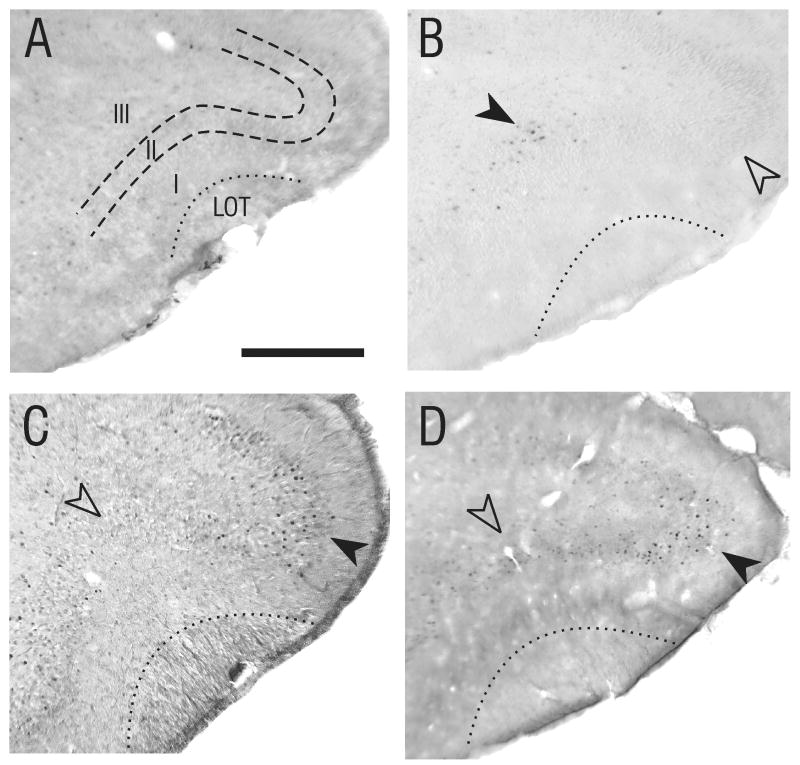
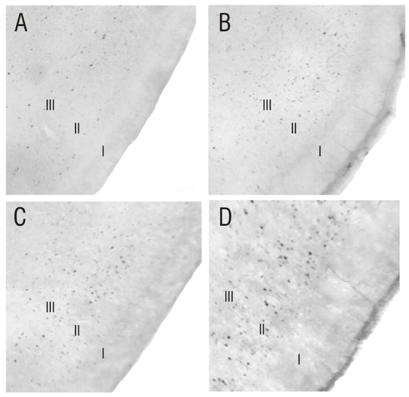
In adult rats, odor-evoked Fos protein expression is found in rostrocaudally-oriented bands of cells in anterior piriform cortex (APC), likely indicating functionally distinct subregions, while activated cells in posterior piriform cortex (PPC) lack apparent spatial organization. To determine whether these patterns are present during early postnatal life, and whether they change during development, Fos expression was assessed following acute exposure to single aliphatic acid odors in developing rats beginning at postnatal day 3 (P3). In the olfactory bulb, Fos-immunoreactive cells were present in the granule cell, mitral cell and glomerular layers at the earliest ages examined. Cells immunopositive for Fos were clustered in areas previously reported as active in response to these odors. In piriform cortex, activation in layers II/III shared some features with that seen in the adult; in APC, rostro-caudally oriented bands of Fos-positive cells alternated with bands relatively free of label, while labeled cells were found dispersed throughout PPC. However, in P3-P7 animals, Fos-positive cells in APC were found in a central rostro-caudally oriented band that was flanked by two bands relatively free of Fos-positive cells. This contrasted with the adult pattern, a central cell-poor band flanked by cell-rich bands, which was observed beginning at P10. These results suggest that subregions of APC visualized by odor-evoked Fos expression are active and functionally distinct shortly after birth. Changes in activity within these subregions during early postnatal development coincide with a shift toward adult-like olfactory learning behavior in the second postnatal week, and may play a role in this behavioral shift.


Figures

Figure 1
Figure 1
Patterns of odor-evoked Fos labeling in the olfactory bulb. A) Coronal section through the olfactory bulb of a P3 rat pup exposed to purified air. D=dorsal; L=Lateral. B) Matched section from a P3 pup (littermate to P3 pup shown in A) exposed to butyric acid. Fos-positive cells were present in the granule cell layer (GCL; solid arrowhead), mitral cell layer (MCL), external plexiform layer (EPL; arrow), and glomerular layer (GL; open arrowhead). Labeling following butyric acid exposure was restricted to the dorsomedial quadrant at all ages tested. These patterns were observed at P5, P7, P10 (C), P20 and P30 (D).
Figure 2
Figure 2
Odor-evoked patterns of Fos labeling in the APC change during development. Fos labeling is found in layers II and III of piriform cortex in rats exposed to butyric acid at all ages tested (P3-P30). However, the spatial distribution of Fos-labeled cells shifted during early postnatal development. A) Coronal section (approximately 3.2mm anterior to bregma) from a P3 animal exposed to purified air only. B) Matched section from a P3 animal (littermate to pup shown in A) exposed to butyric acid. Fos-positive cells were clustered in a location deep to the LOT (solid arrowhead). This pattern also was seen in P5 and P7 animals (not shown). C) At P10, the cluster of Fos-positive cells evoked by butyric acid exposure in APC had shifted dorsally (solid arrowhead), while the area deep to the LOT had become relatively free of Fos-positive cells (open arrowhead). This pattern of labeling continued in animals to P30 (D).
Figure 3
Figure 3
Spatially-distributed patterns of Fos labeling in the PPC do not change during development. Odor-evoked activity was found in layers II and III of PPC at all ages tested. Unlike APC, cellular activity was highly distributed spatially, with no broadscale clustering of Fos-labeled cells apparent. A) Coronal section through the PPC of a P3 animal exposed to purified air, showing minimal Fos expression. B) The number of Fos-positive cells increased following exposure to butyric acid (P3 littermate to pup shown in A). Note the spatially distributed nature of odor-evoked activity in PPC, which was seen at all ages tested, including P10 (C) and P30 (D).
Figure 4
Figure 4
Summary of odor-evoked patterns of Fos labeling in the developing piriform cortex. A) Schematic drawing of the ventrolateral aspect of the rat brain, showing the relative position of the APC and PPC. AON, anterior olfactory nucleus; OB, olfactory bulb; OT, olfactory tubercle; rs, rhinal sulcus. B) Diagram showing the location of highest density of Fos-positive cells following odor exposure within the first postnatal week. Compared with the patterns seen at P10 and later (C), the most notable shift in odor-evoked Fos expression during development was observed in the dorsal band (DB) and central band (CB). Odor-evoked Fos expression in the sulcal band (SB), ventral band (VB) and PPC did not appear to change during development. Scale bar in A = 2 mm.

References

    1. Bolles RC, Woods PJ. The ontogeny of behavior in the albino rat. Animal Behavior. 1965;12:427–441.
    1. Brunjes PC, Illig KR, Meyer EA. A field guide to the anterior olfactory nucleus (cortex) Brain Research Reviews. 2005;50:305–335. - PubMed
    1. Camp LL, Rudy JW. Changes in the categorization of appetitive and aversive events during postnatal development of the rat. Developmental Psychobiology. 1988;21:25–42. - PubMed
    1. Ekstrand JJ, Domroese ME, Feig SL, Illig KR, Haberly LB. Immunocytochemical analysis of basket cells in rat piriform cortex. J Comp Neurol. 2001a;434:308–328. - PubMed
    1. Ekstrand JJ, Domroese ME, Johnson DMG, Feig SL, Knodel SM, Behan M, Haberly LB. A new subdivision of anterior piriform cortex and associated deep nucleus with novel features of interest for olfaction and epilepsy. J Comp Neurol. 2001b;434:289–307. - PubMed

Publication types

MeSH terms

Substances