Morphological and physiological evidence for interstitial cell of Cajal-like cells in the guinea pig gallbladder
- PMID: 17204499
- PMCID: PMC2075389
- DOI: 10.1113/jphysiol.2006.122861
Morphological and physiological evidence for interstitial cell of Cajal-like cells in the guinea pig gallbladder
Abstract
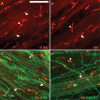
Gallbladder smooth muscle (GBSM) exhibits spontaneous rhythmic electrical activity, but the origin and propagation of this activity are not understood. We used morphological and physiological approaches to determine whether interstitial cells of Cajal (ICC) are present in the guinea pig extrahepatic biliary tree. Light microscopic studies involving Kit tyrosine kinase immunohistochemistry and laser confocal imaging of Ca(2+) transients revealed ICC-like cells in the gallbladder. One type of ICC-like cell had elongated cell bodies with one or two primary processes and was observed mainly along GBSM bundles and nerve fibres. The other type comprised multipolar cells that were located at the origin and intersection of muscle bundles. Electron microscopy revealed ICC-like cells that were rich in mitochondria, caveolae and smooth endoplasmic reticulum and formed close appositions between themselves and with GBSM cells. Rhythmic Ca(2+) flashes, which represent Ca(2+) influx during action potentials, were synchronized in any given GBSM bundle and associated ICC-like cells. Gap junction uncouplers (1-octanol, carbenoxolone, 18beta-glycyrrhetinic acid and connexin mimetic peptide) eliminated or greatly reduced Ca(2+) flashes in GBSM, but they persisted in ICC-like cells, whereas the Kit tyrosine kinase inhibitor, imanitib mesylate, eliminated or reduced action potentials and Ca(2+) flashes in both cell types, as well as associated tissue contractions. This study provides morphological and physiological evidence for the existence of ICC-like cells in the gallbladder and presents data supporting electrical coupling between ICC-like and GBSM cells. The results support a role for ICC-like cells in the generation and propagation of spontaneous rhythmicity, and hence, the excitability of gallbladder.
Figures


Similar articles
-
Spontaneous electrical rhythmicity and the role of the sarcoplasmic reticulum in the excitability of guinea pig gallbladder smooth muscle cells.Am J Physiol Gastrointest Liver Physiol. 2006 Apr;290(4):G655-64. doi: 10.1152/ajpgi.00310.2005. Epub 2005 Nov 17. Am J Physiol Gastrointest Liver Physiol. 2006. PMID: 16293655
-
Distribution of the interstitial Cajal-like cells in the gallbladder and extrahepatic biliary duct of the guinea-pig.Acta Histochem. 2009;111(2):157-65. doi: 10.1016/j.acthis.2008.05.005. Epub 2008 Aug 3. Acta Histochem. 2009. PMID: 18676008
-
Role of mitochondria in spontaneous rhythmic activity and intracellular calcium waves in the guinea pig gallbladder smooth muscle.Am J Physiol Gastrointest Liver Physiol. 2008 Feb;294(2):G467-76. doi: 10.1152/ajpgi.00415.2007. Epub 2007 Nov 29. Am J Physiol Gastrointest Liver Physiol. 2008. PMID: 18048480
-
Smooth muscle cells, interstitial cells and neurons in the gallbladder (GB): Functional syncytium of electrical rhythmicity and GB motility (Review).Int J Mol Med. 2023 Apr;51(4):33. doi: 10.3892/ijmm.2023.5236. Epub 2023 Mar 17. Int J Mol Med. 2023. PMID: 36928163 Free PMC article. Review.
-
Structure and organization of interstitial cells of Cajal in the gastrointestinal tract.J Physiol. 2006 Nov 1;576(Pt 3):653-8. doi: 10.1113/jphysiol.2006.116624. Epub 2006 Aug 17. J Physiol. 2006. PMID: 16916909 Free PMC article. Review.
Cited by
-
Distribution changes of interstitial cells of Cajal during cholesterol gallstone formation in guinea pigs fed a high cholesterol diet.Int J Clin Exp Pathol. 2018 Mar 1;11(3):1653-1659. eCollection 2018. Int J Clin Exp Pathol. 2018. PMID: 31938265 Free PMC article.
-
A distinct type of cell in myocardium: interstitial Cajal-like cells (ICLCs).J Cell Mol Med. 2009 Feb;13(2):295-308. doi: 10.1111/j.1582-4934.2008.00668.x. Epub 2009 Jan 27. J Cell Mol Med. 2009. PMID: 19183408 Free PMC article.
-
Serotonin augments gut pacemaker activity via 5-HT3 receptors.PLoS One. 2011;6(9):e24928. doi: 10.1371/journal.pone.0024928. Epub 2011 Sep 15. PLoS One. 2011. PMID: 21949791 Free PMC article.
-
Morphological expression of KIT positive interstitial cells of Cajal in human bladder.J Urol. 2010 Jul;184(1):370-7. doi: 10.1016/j.juro.2010.03.005. Epub 2010 May 20. J Urol. 2010. PMID: 20488490 Free PMC article.
-
Effect of cholesterol on in vitro cultured interstitial Cajal-like cells isolated from guinea pig gallbladders.World J Gastrointest Surg. 2020 May 27;12(5):226-235. doi: 10.4240/wjgs.v12.i5.226. World J Gastrointest Surg. 2020. PMID: 32551028 Free PMC article.
References
-
- Balemba OB, Heppner TJ, Bonev AD, Nelson MT, Mawe GM. Calcium waves in intact guinea pig gallbladder smooth muscle cells. Am J Physiol Gastrointest Liver Physiol. 2006 a;291:G717–G727. - PubMed
-
- Balemba OB, Salter MJ, Heppner TJ, Bonev AD, Nelson MT, Mawe GM. Spontaneous electrical rhythmicity and the role of the sarcoplasmic reticulum in the excitability of guinea pig gallbladder smooth muscle cells. Am J Physiol Gastrointest Liver Physiol. 2006 b;290:G655–G664. - PubMed
-
- Biers SM, Reynard JM, Doore T, Brading AF. The functional effects of a c-kit tyrosine inhibitor on guinea-pig and human detrusor. BJU Int. 2006;97:612–616. - PubMed
-
- Burns AJ, Herbert TM, Ward SM, Sanders KM. Interstitial cells of Cajal in the guinea-pig gastrointestinal tract as revealed by c-Kit immunohistochemistry. Cell Tissue Res. 1997;290:11–20. - PubMed
Publication types
MeSH terms
Substances
Grants and funding
LinkOut - more resources
Full Text Sources
Miscellaneous

