. 2007 Jan 16;104(3):1021-6.
doi: 10.1073/pnas.0610294104. Epub 2007 Jan 4.

A natural genetic code expansion cassette enables transmissible biosynthesis and genetic encoding of pyrrolysine

Affiliations

A natural genetic code expansion cassette enables transmissible biosynthesis and genetic encoding of pyrrolysine

David G Longstaff et al. Proc Natl Acad Sci U S A. .

Abstract

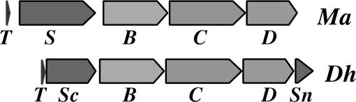
Pyrrolysine has entered natural genetic codes by the translation of UAG, a canonical stop codon. UAG translation as pyrrolysine requires the pylT gene product, an amber-decoding tRNA(Pyl) that is aminoacylated with pyrrolysine by the pyrrolysyl-tRNA synthetase produced from the pylS gene. The pylTS genes form a gene cluster with pylBCD, whose functions have not been investigated. The pylTSBCD gene order is maintained not only in methanogenic Archaea but also in a distantly related Gram-positive Bacterium, indicating past horizontal gene transfer of all five genes. Here we show that lateral transfer of pylTSBCD introduces biosynthesis and genetic encoding of pyrrolysine into a naïve organism. PylS-based assays demonstrated that pyrrolysine was biosynthesized in Escherichia coli expressing pylBCD from Methanosarcina acetivorans. Production of pyrrolysine did not require tRNA(Pyl) or PylS. However, when pylTSBCD were coexpressed with mtmB1, encoding the methanogen monomethylamine methyltransferase, UAG was translated as pyrrolysine to produce recombinant monomethylamine methyltransferase. Expression of pylTSBCD also suppressed an amber codon introduced into the E. coli uidA gene. Strains lacking one of the pylBCD genes did not produce pyrrolysine or translate UAG as pyrrolysine. These results indicated that pylBCD gene products biosynthesize pyrrolysine using metabolites common to Bacteria and Archaea and, furthermore, that the pyl gene cluster represents a "genetic code expansion cassette," previously unprecedented in natural organisms, whose transfer allows an existing codon to be translated as a novel endogenously synthesized free amino acid. Analogous cassettes may have served similar functions for other amino acids during the evolutionary expansion of the canonical genetic code.


Conflict of interest statement

The authors declare no conflict of interest.

Figures

Fig. 1.
Fig. 1.
The pyl genes from the methanogenic archaeon M. acetivorans (Ma) and the Gram-positive Bacterium D. hafniense (Dh). The gene order of the pyl genetic code expansion cassette is conserved, with the exception that the D. hafniense pylS gene homolog has been split into two genes encoding homologs to the PylS N-terminal domain (pylSn) and the catalytic core domain (pylSc) that now flank pylBCD (7).
Fig. 2.
Fig. 2.
UAG translation in mtmB1 in E. coli bearing the five pyl genes. SDS-solubilized extracts of E. coli transformed with pylT, pylS, and mtmB1 on pDLBAD were analyzed by anti-MtmB1 immunoblot. Additionally, the strains were also transformed with the following: lane 1, the vector pACYCDuet-1; lane 2, pK13 bearing pylB, pylC, and pylD; lane 3, pK14 bearing pylB and pylD; lane 4, pK15 bearing pylC and pylD; and lane 5, pK16 bearing pylB and pylC. Lane 6 contains a set of standard proteins with molecular masses indicated at the right in kilodaltons, whereas lane 7 contains purified MtmB1. The upper arrow indicates the location of the mtmB1 UAG translation product, and the lower arrow indicates the location of the mtmB1 UAG termination product.
Fig. 3.
Fig. 3.
Cells bearing pylB, pylC, and pylD produce pyrrolysine detectable by in vitro assays using pyrrolysyl-tRNA synthetase. (A) The direct aminoacylation of tRNAPyl with pyrrolysine present in cell extracts by PylS. Charged and uncharged tRNA species in the isolated cellular pool of tRNAPyl were separated in an acid-urea polyacrylamide gel that was subsequently electroblotted. The blot was then probed with 32P-labeled oligodeoxynucleotide complementary to tRNAPyl and analyzed by phosphorimager. Lane 1 was loaded with cellular tRNA as isolated (9) and has both charged (upper arrow) and uncharged (lower arrow) tRNAPyl, whereas lane 2 is the cellular tRNA pool following deacylation at pH 9 for 30 min and shows only uncharged tRNAPyl. Aminoacylation of tRNAPyl by PylS with pyrrolysine was then tested in 25-μl reactions that contained: 3.2 μM PylS, 50 mM KCl, 1 mM MgCl2, 5 mM ATP, 0.5 mM DTT, 8 μg of M. acetivorans deacylated cellular tRNA, and the metabolite pool from the indicated E. coli strains in 10 mM Hepes buffer, pH 7.2. After incubation for 40 min at 37°C, aminoacylation was tested as above in reactions that also contained the following: lane 3, no metabolite pool; or the pool from, lane 4, pK13 bearing pylB, pylC, and pylD; 5, pK14 bearing pylB and pylD; 6, pK15 bearing pylC and pylD; 7, pK16 bearing pylB and pylC; and 8, pACYCDuet-1 bearing no pyl gene. (B) Cellular amino acid pools were tested for activity in the pyrophosphate:ATP exchange assay mediated by PylS in the presence of pyrrolysine. Exchange of 32P-pyrophosphate into ATP was monitored in 100-μl reactions containing 5.5 μM PylS, 10 mM MgCl2, 25 mM KCl, 1 mM potassium fluoride, 4 mM DTT, 2 mM ATP, and 2 mM 32P-PPi (12 dpm/pmol) in 20 mM Hepes-KOH (pH 7.2) and incubated at 37°C. Aliquots were removed at the time points indicated, and the amount of radiolabel bound to acid-washed activated charcoal was quantified to estimate the amount of 32ATP formed. Shown are illustrated results from averaged duplicate reactions that were supplemented with the extracted amino acid pool from pK13 (●), pK14 (X), pK15 (△), pK16 (◇), or pACYCDuet-1 (○).
Fig. 4.
Fig. 4.
Mass spectrometry demonstrates that the UAG-encoded residue of recombinant mtmB1 is pyrrolysine in cells bearing the five pyl genes. (Inset) An anti-MtmB1 immunoblot of the 7-M urea-solubilized fraction from the cell pellet of French-press-lysed cells transformed with pDLBAD and either pACYCDuet-1 or pK13, as indicated. The upper arrow points to the UAG-translation mtmB1 product, whereas the lower arrow points to the UAG-termination product. Subsequently, the 7-M urea fraction was electrophoresed in an SDS-polyacrylamide gel, and the 50-kDa MtmB1 band was excised and subjected to in-gel digestion with chymotrypsin and analysis by mass spectrometry. The predicted sequence of the peptide ion at m/z = 783.432+ from chymotryptic cleavage of MtmB1 is shown along with the fragmentation sites forming the a-, b-, and y-series ions identified in the ion's collision-induced dissociation spectrum shown below. The peak of the parent ion (PI) is indicated. A horizontal arrow lies between the a-, b-, or y-series ion pairs whose mass differences allow calculation of the mass of the pyrrolysyl residue. The masses of the remaining identified ion peaks are listed in SI Table 1.

Similar articles

Cited by

References

    1. Paul L, Ferguson DJ, Krzycki JA. J Bacteriol. 2000;182:2520–2529. - PMC - PubMed
    1. Burke SA, Lo SL, Krzycki JA. J Bacteriol. 1998;180:3432–3440. - PMC - PubMed
    1. James CM, Ferguson TK, Leykam JF, Krzycki JA. J Biol Chem. 2001;276:34252–34258. - PubMed
    1. Hao B, Gong W, Ferguson TK, James CM, Krzycki JA, Chan MK. Science. 2002;296:1462–1466. - PubMed
    1. Hao B, Zhao G, Kang P, Soares J, Ferguson T, Gallucci J, Krzycki J, Chan M. Chem Biol. 2004;11:1317–1324. - PubMed

Publication types

MeSH terms