. 2006 Dec 27;1(1):e106.
doi: 10.1371/journal.pone.0000106.

An NF-kappaB and slug regulatory loop active in early vertebrate mesoderm

Affiliations

An NF-kappaB and slug regulatory loop active in early vertebrate mesoderm

Chi Zhang et al. PLoS One. .

Abstract

Background: In both Drosophila and the mouse, the zinc finger transcription factor Snail is required for mesoderm formation; its vertebrate paralog Slug (Snai2) appears to be required for neural crest formation in the chick and the clawed frog Xenopus laevis. Both Slug and Snail act to induce epithelial to mesenchymal transition (EMT) and to suppress apoptosis.

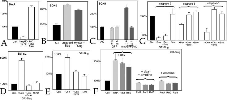
Methodology & principle findings: Morpholino-based loss of function studies indicate that Slug is required for the normal expression of both mesodermal and neural crest markers in X. laevis. Both phenotypes are rescued by injection of RNA encoding the anti-apoptotic protein Bcl-xL; Bcl-xL's effects are dependent upon IkappaB kinase-mediated activation of the bipartite transcription factor NF-kappaB. NF-kappaB, in turn, directly up-regulates levels of Slug and Snail RNAs. Slug indirectly up-regulates levels of RNAs encoding the NF-kappaB subunit proteins RelA, Rel2, and Rel3, and directly down-regulates levels of the pro-apopotic Caspase-9 RNA.

Conclusions/significance: These studies reveal a Slug/Snail-NF-kappaB regulatory circuit, analogous to that present in the early Drosophila embryo, active during mesodermal formation in Xenopus. This is a regulatory interaction of significance both in development and in the course of inflammatory and metastatic disease.


Conflict of interest statement

Competing Interests: The authors have declared that no competing interests exist.

Figures

Figure 1
Figure 1. Slug morpholino effects:
Panel A is a comparison of the Slug morpholino sequence (“MO”) with X. laevis SlugA, SlugB, and Snail RNA sequences; start codons are underlined. B–G: Injection of the Slug MO (10 ng/embryo) blocks the expression of Xbra (B-uninjected, C-Slug MO injected), Xmenf (D-uninjected, E-injected), and Antipodean (Apod)(F-uninjected, G-injected). Arrows (C,E,G) point to region of suppression; vegetal pole (“VP”) is indicated. In G, the red staining is due to a β-galactosidase lineage marker. H: Injection of the Slug MO into one cell of a two cell embryo blocks the expression of Sox9 on the injected side (arrow); “*” marks otic placode domain of Sox9 expression. I: Sox9 expression in Slug MO injected embryos is rescued by co-injection of mycGFP-Slug RNA (650 pg/embryo). In analogous studies, the effects of the Slug MO (10 ng/embryo) on Xbra (J), Apod (K) and Sox9 (L) expression were rescued by injection of Snail RNA (500 pg/embryo). In H, I and L, the line marks midline of the embryo, with anterior (“An”) and posterior (“Ps”) indicated.
Figure 2
Figure 2. Timing of Slug rescue of Slug MO phenotypes:
To analyze the timing of Slug activity in the early embryo, we injected one cell of two cell embryos with Slug MO (10 ng/embryo) together with RNA (650 pg/embryo) encoding the chimeric GR-Slug-GFP protein. A: In the absence of the activating drug dexamethasone, the Slug MO phenotype, i.e. suppression of Xbra expression in stage 11 embryos (A)(arrow) and suppression of Sox9 expression in stage 16 embryos (D), was unaltered. When embryos were treated with dexamethasone (20 µM) beginning at stage 8, there as an essentially complete rescue of Xbra (B,C) and Sox9 expression (E,H). Treatment of embryos with dexamethasone at stage 11 (early gastrulation) was also effective at rescuing Sox9 expression (F,H), while addition of dexamethasone at stage 13 (late gastrulation/early neurulation) produced at most a partial and inefficient rescue of Sox9 expression (G,H).
Figure 3
Figure 3. Rescue of Slug MO effects by Bcl-xL
A: Injection of the Slug MO leads to an increase in TUNEL staining. B: This increase is blocked by the injection of Bcl-xL RNA (600 pg/embryo and co-injected with LacZ RNA). Injected sides of embryos are marked by an “*” and red staining; line marks midline of the embryo, with anterior (“An”) and posterior (“Ps”) indicated. Injection of Bcl-xL RNA (600 pg/embryo) rescues Apod (C- Slug MO injected, D-Slug MO+Bcl-xl RNA injected), Xbra (Ε- Slug MO injected, F-Slug MO+Bcl-xl RNA injected), and Sox9 expression (G- Slug MO injected, H-Slug MO+Bcl-xL RNA injected). I: Injection of Bcl-xL RNA into one cell of a two-cell embryo led to a dramatic increase in the intensity and extent of Slug expression at stage 16; the region of Slug expression on the uninjected (control) side of the embryo is indicated by the dashed circle. J: Injection of Bcl-xL RNA produced an increase in Slug RNA levels in animal caps prepared at stage 8/9 and analyzed by QRT-PCR when uninjected embryos reached stage 11. Ornithine decarboxylase (ODC) was used as normalization control. RNA levels in the control case were set to 100%.
Figure 4
Figure 4. Characterization of Bcl-xL effects on NF-κB activity.
A: Fertilized eggs were injected with RNA (650 pg/embryo) encoding Xenopus IκBα-V5. Beginning at stage 8, experimental embryos were treated with 50 µM AKBA and analyzed at stage 11 by SDS-PAGE/immunoblot using an anti-V5 antibody and the antiSOX3c antibody to visualize endogenous Sox3 protein as a loading control. AKBA treatment stabilized the IκBα-V5 polypeptide. B: Fertilized eggs were injected with p3XκB-firefly luciferase (“3κB-Luc”) and pTK-Renilla luciferase (“RL-TK”) DNAs (10 pg/embryo each) either alone (“Con”) or together with Bcl-xL (500 pg/embryo) RNA, or Bcl-xL and IκBsa (600 pg/embryo) RNAs. Alternatively, animal caps prepared from Bcl-xL RNA injected embryos were cultured in either control buffer (0.1% DMSO), 20 µM or 50 µM AKBA. At stage 11, caps were analyzed for luciferase activity. Bcl-xL induced an increase in 3XκB-Luc activity that was blocked by either IκBsa or AKBA. Error bars in B reflect standard deviation from the mean of multiple experiments.
Figure 5
Figure 5. Bcl-xL regulation of NF-κB RNAs:
A: RNA was extracted from eggs and embryos at various stages and analyzed by RT-PCR (28 cycles); levels of RelA, Rel2, Rel3, RelB and Xp100 RNAs drop between stage 7 and 9 and, except for RelB, increase following gastrulation (stage 12/13). Levels of Bcl-xL RNA appear relatively constant throughout this period of development. B: At stage 16, embryos were dissected into anterior dorsal (AD), posterior dorsal (PD), anterior ventral (AV), and posterior ventral (PV) quadrants and RNA was analyzed by RT-PCR; RelB was not expressed at this stage; expression of RelA, Slug and Sox9 are restricted to the anterior dorsal quadrant, while Bcl-xL, Rel2, Rel3, and Xp100 RNAs can be detected throughout the embryo. C, D: Animal caps were prepared from embryos injected with GR-Bcl-xL-GFP RNA (“GRBclxL”)(600 pg/embryo) and either left untreated (0.1%DMSO)(“Con”), treated with 20 µM dexamethasone (“+Dex”), treated first with 100 µg/ml emetine and then dexamethasone (“+Dex+Eme”), or treated with emetine alone (“+Eme”), and analyzed at stage 11 for Slug, RelA (C), Rel2 and Rel3 (D) RNA levels. Treatment with emetine blocked the increase in Slug and Rel2, but not RelA and Rel3 RNAs; emetine treatment alone produced control or slightly reduced levels of Slug and Rel RNAs. E: Activation of the GR-Bcl-xL-GFP protein in embryos injected with the Slug MO produces an increase in the level of Snail RNA, analyzed at stage 11. F: In animal caps derived from GR-BclxL-GFP RNA injected embryos, AKBA (50 µM) inhibited the dexamethasone-induced increases in Slug, Snail, and RelA RNA levels; while treatment with AKBA alone lead to a decrease in Slug, Snail and RelA RNA levels. G: In animal caps, injection of RelAΔSP RNA (600 pg/embryo) blocked Rel3 and Xp52 RNA induced activation of the 3XκB reporter. H: The ability of Bcl-xL RNA to increase levels of RelA and Slug RNAs in animal caps was blocked by the co-injection of RelAΔSP RNA. Error bars in C–H reflect standard deviation from the mean of multiple experiments.
Figure 6
Figure 6. NF-κB regulation of mesodermal and neural markers:
The Slug MO induced loss of Xbra (A,B), Apod (C,D) and Sox9 (E,F) expression was rescued by injection of RelA RNA (600 pg/embryo)(A, C, E-Slug MO alone, B, D, F-Slug MO+RelA RNA). G: Treatment of early embryos with AKBA (50 µM from the 4-cell stage on) lead to a decrease in Xbra staining (control and AKBA-treated embryos marked). Injection of RNA encoding IκBsa (H,I) or RelAΔSP (J–L) had effects similar to that seen in Slug MO injected embryos; that is, both induced the reduction of Xbra (H,J), Apod (K) and Sox9 (I,L) RNA staining. AKBA treatment had no reproducible effect on Sox9 expression (data not shown). Arrows mark affected regions.
Figure 7
Figure 7. NF-κB's regulatory targets:
A: In animal caps, RelA lead to an increase in Slug RNA levels, while RelAΔSP produced a decrease. When activated by dexamethasone (+Dex), the hormone-regulated form of RelA, GR-RelA (600 pg RNA/embryo), induced a similar increase in the levels of Slug RNA, as well as Snail, Sox9 (B), and Bcl-xL (C) RNAs compared to animal caps from GR-RelA injected embryos not exposed to dexamethasone. Similar effects were seen in the presence of emetine (+Dex+Eme,), while emetine alone (+Eme) had little effect on any of measured RNA levels. Error bars in reflect standard deviation from the mean of multiple experiments.
Figure 8
Figure 8. Slug's regulatory targets:
A: In animal caps analyzed at stage 11, the Slug MO (10 ng/embryo) produced a decrease in RelA RNA levels that was rescued by co-injection of mycGFP-Slug RNA (1 ng/embryo). B: Animal caps were prepared from embryos injected with either untagged or mycGFP-Slug RNAs; both produced a similar increase in Sox9 RNA levels. C: Animal caps, from embryos injected with either mt-GFP or mycGFP-Slug RNAs, were analyzed when control embryos reached stage 11 or stage 16; at stage 11 mycGFP-Slug induced an increase in Sox9 RNA levels, which returned to baseline by stage 16. No change in Sox9 RNA levels were observed at either stage in animal caps expressing mt-GFP. In GR-Slug injected caps, levels of Bcl-xL (D), Sox9 (E), RelA, Rel2 and Rel3 (F), and caspase-9, -3 and -6 (G) RNAs were increased in response to dexamethasone; with the sole exception of caspase-9, these increases were blocked by emetine. In all panels, error bars reflect standard deviation from the mean of multiple experiments.
Figure 9
Figure 9. Bcl-xL-Slug-NF-κB network:
This diagram focuses on the regulatory interactions uncovered in the course of our studies (see text for caveats associated with the identification of direct interactions). Protein names are underlined, gene names are in italics. Bcl-xL appears to activate NF-κB through effects on IκK activity and IκB stability. NF-κB acts directly to regulate Slug, Snail, RelA, and Rel3 levels; NF-κB regulation of the expression of its inhibitor IκB is based on data from mammalian systems. Caspase-9 was the only direct target of Slug identified in our studies; indirect interactions are indicated by dotted lines.

Similar articles

Cited by

References

    1. Thiery JP, Sleeman JP. Complex networks orchestrate epithelial-mesenchymal transitions. Nat Rev Mol Cell Biol. 2006;7:131–42. - PubMed
    1. Fritzenwanker JH, Saina M, Technau U. Analysis of forkhead and snail expression reveals epithelial-mesenchymal transitions during embryonic and larval development of Nematostella vectensis. Dev Biol. 2004;275:389–402. - PubMed
    1. Technau U, Scholz CB. Origin and evolution of endoderm and mesoderm. Int J Dev Biol. 2003;47:531–9. - PubMed
    1. Vickaryous MK, Hall BK. Human cell type diversity, evolution, development, and classification with special reference to cells derived from the neural crest. Biol Rev Camb Philos Soc. 2006;81:425–55. - PubMed
    1. Whiteley M, Noguchi PD, Sensabaugh SM, Odenwald WF, Kassis JA. The Drosophila gene escargot encodes a zinc finger motif found in snail-related genes. Mech Dev. 1992;36:117–27. - PubMed

Publication types

MeSH terms