. 2006 Dec 27;1(1):e119.
doi: 10.1371/journal.pone.0000119.

Designer self-assembling peptide nanofiber scaffolds for adult mouse neural stem cell 3-dimensional cultures

Affiliations

Designer self-assembling peptide nanofiber scaffolds for adult mouse neural stem cell 3-dimensional cultures

Fabrizio Gelain et al. PLoS One. .

Abstract

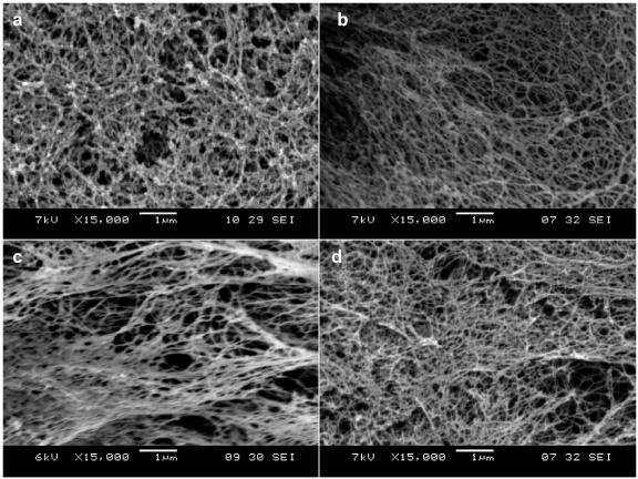
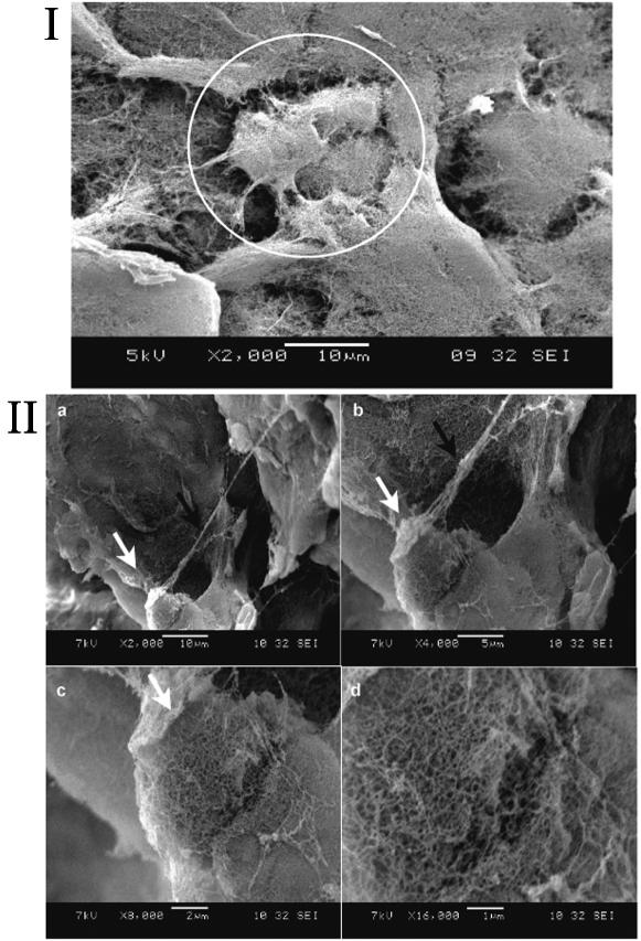
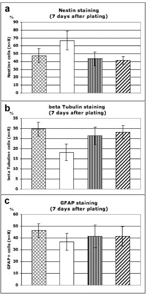
Biomedical researchers have become increasingly aware of the limitations of conventional 2-dimensional tissue cell culture systems, including coated Petri dishes, multi-well plates and slides, to fully address many critical issues in cell biology, cancer biology and neurobiology, such as the 3-D microenvironment, 3-D gradient diffusion, 3-D cell migration and 3-D cell-cell contact interactions. In order to fully understand how cells behave in the 3-D body, it is important to develop a well-controlled 3-D cell culture system where every single ingredient is known. Here we report the development of a 3-D cell culture system using a designer peptide nanofiber scaffold with mouse adult neural stem cells. We attached several functional motifs, including cell adhesion, differentiation and bone marrow homing motifs, to a self-assembling peptide RADA16 (Ac-RADARADARADARADA-COHN2). These functionalized peptides undergo self-assembly into a nanofiber structure similar to Matrigel. During cell culture, the cells were fully embedded in the 3-D environment of the scaffold. Two of the peptide scaffolds containing bone marrow homing motifs significantly enhanced the neural cell survival without extra soluble growth and neurotrophic factors to the routine cell culture media. In these designer scaffolds, the cell populations with beta-Tubulin(+), GFAP(+) and Nestin(+) markers are similar to those found in cell populations cultured on Matrigel. The gene expression profiling array experiments showed selective gene expression, possibly involved in neural stem cell adhesion and differentiation. Because the synthetic peptides are intrinsically pure and a number of desired function cellular motifs are easy to incorporate, these designer peptide nanofiber scaffolds provide a promising controlled 3-D culture system for diverse tissue cells, and are useful as well for general molecular and cell biology.


Conflict of interest statement

Competing Interests: SZ is one of the inventors of the self-assembling peptides and also a co-founder of 3DM, Inc., an MIT startup that licenses the peptide scaffold patents to BD Biosciences for distribution. SZ also receives free samples of RADA16, peptide scaffold materials without the addition of functional motifs from 3DM as the controls.

Figures

Figure 1
Figure 1
Molecular and schematic models of the designer peptides and of the scaffolds. a) Molecular models of RADA16, RADA16-Bone Marrow Homing Peptide 1 (BMHP1) and RADA16-Bone Marrow Homing Peptide 2 (BMHP2). RADA16 is an alternating16-residue peptide with basic arginine (blue), hydrophobic alanine (white) and aspartic acid (red). These peptides self-assemble once exposed to physiological pH solutions or salt. The alanines of the RADA16 providing hydrophobic interaction are on one side of the peptide, and the arginines and aspartates form complementary ionic bonds on the other. The BMHP1 and BMHP2 motifs were directly extended from RADA16 with two glycine spacers and are composed of a lysine (blue), serine and threonine (green) and different hydrophobic (white) residues. Neutral polar residues are drawn in green. b) Schematic models of several different functional motifs (different colored bars) could be extended from RADA16 (blue bars) in order to design different peptides (I, II, III, IV and V). They can be combined in different ratios. A schematic model of a self-assembling nanofiber scaffold with combinatorial motifs carrying different biological functions is shown.
Figure 2
Figure 2
SEM images of Matrigel and various designer peptide nanofiber scaffolds. a) Matrigel, b) RADA16, c) RADA16-BMHP1, d) RADA16-BMHP2 nanofiber scaffolds assembled in PBS solutions. Matrigel nanostructures are comparable in size to nanofibers found after self-assembly of the designer peptides. Clusters and aggregates of the unidentified naturally derived proteins in Matrigel (a) are absent in the pure peptide scaffolds shown in (b), (c) and (d). The interwoven nanofibers are ∼10 nm in diameter in each of the peptide scaffolds with ∼5–200 nm pores. The appended functional motifs did not prevent peptide self-assembly.
Figure 3
Figure 3
SEM images of adult mouse neural stem cells (NSC) embedded in designer peptide nanofiber scaffold RADA16-BMHP1 (1% v/w) after 14 day in vitro cultures. I) Cluster of three visible NSCs (white circle) embedded in 3-D self-assembling RADA16-BMHP1. II) A single cell at different magnification with extended processes embedded in the scaffold is shown (a–c). White arrows point to the image areas enlarged in the consecutive pictures. d) High-magnification picture focusing on the interface between the nanofiber scaffold and the round shaped cell body. The black arrow in (b) points to a cellular process. Cells and processes are thus embedded in the self-assembling peptide nanofiber scaffold in a true 3-D environment, which may likely promote cell adhesions in 3-D similar to the natural cellular environment. Adult mouse neural stem cells have been cultured and could be differentiated in vitro for several weeks. The scale bars are shown on each image.
Figure 4
Figure 4
MTT cell proliferation assays of adult mouse neural stem cells after 7-day culture. Cells were seeded on Matrigel or peptide scaffolds. The results are expressed as cell % increases from the seeding population on first day. BMHP1 and BMHP2 peptide scaffolds allow for higher cell proliferation in comparison to RADA16 peptide scaffold (t = −7.28 and t = −5.28 for respectively RADA16 vs. RADA16-BMHP1 and RADA16 vs. RADA16-BMHP2 with p<0.0001% in both cases). Not surprisingly, Matrigel containing various unknown quantity of growth factors showed considerable cell population increase. Similar increases of total cell populations were confirmed for 14-day cultures (not shown). The cell proliferation using the designer peptide scaffolds could be further improved from addition of soluble neurotrophic factors.
Figure 5
Figure 5
Images of neural stem cells in differentiation assays. a–d) Differentiating adult mouse neural stem cells cultured for 7 days in vitro on (a) 1% Matrigel, (b) RADA16 peptide scaffold; (c) RADA16-BMHP1 and (d) RADA16-BMHP2, 1% peptide scaffolds were examined with staining assays for DAPI (cell nuclei in blue), β-Tubulin+ (neurons in red), and Nestin+ (neural progenitors in green). β-Tubulin+ cells on BMHP1 and BMHP2 showed increased branching in comparison with RADA16 scaffold. These appearances are comparable with neurons on Matrigel coated wells. Nestin+ and β-Tubulin+ signals show negligible cross-reaction (MERGE images).
Figure 6
Figure 6
Quantitative cell differentiation assays. The adult mouse neural stem cells were stained with various markers 7 days after culturing on Matrigel (positive control), RADA16, RADA16-BMHP1 and RADA16-BMHP2 peptide scaffolds. Values are expressed as averages±STD. a) % Nestin+ progenitor cell was reduced on the cultures of Matrigel, RADA16-BMHP1 and RADA16-BMHP2 peptide scaffolds, suggesting cell differentiation. b) % β-Tubulin+ cell was similar for Matrigel culture, RADA16-BMHP1 and RADA16-BMHP2 peptide scaffolds, suggesting neuronal differentiation. c) % GFAP+ cell was slightly higher in Matrigel culture, RADA16-BMHP1 and RADA16-BMHP2 peptide scaffolds, suggesting the ability of glial cells to adapt to a wide range of substrates.
Figure 7
Figure 7
Comparisons of the gene expression profiling. Adamts2–5: disintegrin-like and metalloprotease (reprolysin type) with thrombospondin type 1 motif, 2 or 5; Col3a1: Procollagen, type III, alpha 1; Col4a3: Procollagen, type IV, alpha 3; Col5a1: Procollagen, type V, alpha 1; Col5a1: Procollagen, type V, alpha 1; Emilin1: Elastin microfibril interfacer 1; Fbln1: Fibulin 1; Lamb2: Laminin, beta 2; Ncam2: Neural cell adhesion molecule 2; Spock1: Sparc/osteonectin, cwcv and kazal-like domains; Tnc: Tenascin C.

References

    1. Lanza R, Langer R, Vacanti J. Principles of Tissue Engineering, Academic Press; San Diego, USA: 2000. 2nd.
    1. Kleinman HK, McGarvey ML, Hassell JR, Star VL, Cannon FB, et al. Basement membrane complexes with biological activity. Biochemistry. 1986;25:312–318. - PubMed
    1. Weaver VM, Howlett AR, Langton-Webster B, Petersen OW, Bissell MJ. The development of a functionally relevant cell culture model of progressive human breast cancer. Seminar in Cancer Biol. 1995;6:175–184. - PubMed
    1. Spancake KM, Anderson CB, Weaver VM, Matsunami N, Bissell MJ, et al. E7-transduced human breast epithelial cells show partial differentiation in three-dimensional culture. Cancer Res. 1999;59:6042–6045. - PubMed
    1. Zhau HE, Goodwin TJ, Chang SM, Baker TL, Chung LW. Establishment of a three-dimensional human prostate organoid coculture under microgravity-simulated conditions: evaluation of androgen-induced growth and PSA expression. Cell Dev. Biol. Anim. 1997;33:375–380. - PubMed

Publication types

MeSH terms