. 2006 Dec 27;1(1):e126.
doi: 10.1371/journal.pone.0000126.

Docking of secretory vesicles is syntaxin dependent

Affiliations

Docking of secretory vesicles is syntaxin dependent

Heidi de Wit et al. PLoS One. .

Abstract

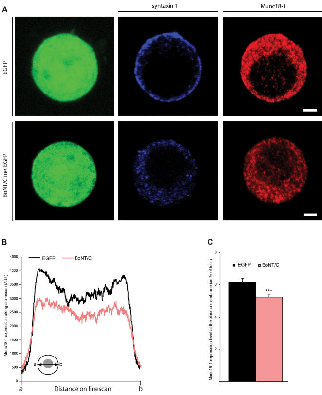
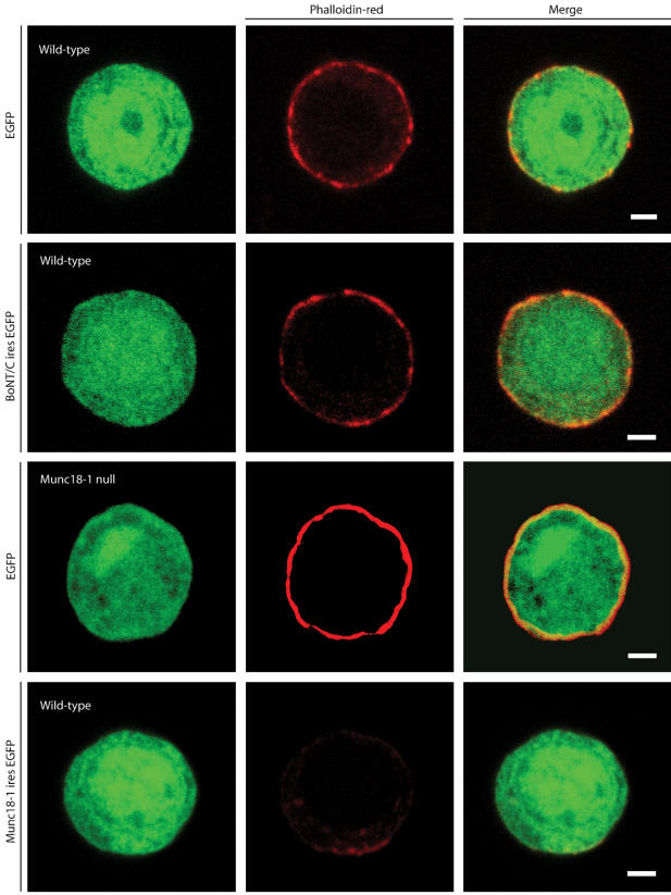
Secretory vesicles dock at the plasma membrane before they undergo fusion. Molecular docking mechanisms are poorly defined but believed to be independent of SNARE proteins. Here, we challenged this hypothesis by acute deletion of the target SNARE, syntaxin, in vertebrate neurons and neuroendocrine cells. Deletion resulted in fusion arrest in both systems. No docking defects were observed in synapses, in line with previous observations. However, a drastic reduction in morphologically docked secretory vesicles was observed in chromaffin cells. Syntaxin-deficient chromaffin cells showed a small reduction in total and plasma membrane staining for the docking factor Munc18-1, which appears insufficient to explain the drastic reduction in docking. The sub-membrane cortical actin network was unaffected by syntaxin deletion. These observations expose a docking role for syntaxin in the neuroendocrine system. Additional layers of regulation may have evolved to make syntaxin redundant for docking in highly specialized systems like synaptic active zones.


Conflict of interest statement

Competing Interests: The authors have declared that no competing interests exist.

Figures

Figure 1
Figure 1
Evoked catecholeamine release is absent in syntaxin deleted chromaffin cells. (A) Fluorescent image of cultured chromaffin cells incubated with SFV-egfp or SFV BoNT/C-ires-egfp, and immunostained for syntaxin, showing a reduced syntaxin staining at the plasma membrane after BoNT/C expression. The syntaxin staining after BoNT/C was slightly overexposed to emphasize the persistence of cytosolic staining (as opposed to plasma membrane staining). Scale bars represent 2 µm. (B) Examples of amperometric recordings in control and BoNT/C infected chromaffin cells during stimulation with a 30 mM K+ solution.
Figure 2
Figure 2
Syntaxin deletion decreases the number of morphologically docked secretory vesicles. (A) Electron micrographs of control and BoNT/C expressing chromaffin cell. For each cell a magnification of a sub-membrane region is shown indicating severely impaired vesicle docking after acute BoNT/C expression compared to the control cell that contains many morphologically docked vesicles at the plasma membrane. Scale bars represent 200 nm. (B) Normalized cumulative distribution of secretory vesicles as a function of distance from the plasma membrane in control cells expressing EFGP or BoNT/C. Inset shows cumulative vesicle distribution in the sub-membrane region within 0–100 nm. Grey line represents the vesicle distribution in the absence of Munc18-1 as shown before . (C–E) Number of docked vesicles (C), vesicles>0–30 and within 30–100 nm (D), and the total number of vesicles (E). Data are mean±SEM from the following number of cells (n) and animals (N): control+EGFP, n = 20, N = 4; control+BoNT/C, n = 20, N = 4 (**p<0.05 and ***p<0.001, ANOVA and student's t-test).
Figure 3
Figure 3
Spontaneous and evoked vesicle fusion is impaired in synapses lacking syntaxin. (A) Representative traces of mEPSC's in whole-cell voltage clamp recordings from control synapses showed frequent spontaneous miniature events, while syntaxin deleted synapses show a strong reduction of spontaneous release. (B) Frequency of spontaneous synaptic events. Numbers indicate mean±SEM for control (n = 5) and BoNT/C infected (n = 4) neurons from N = 2 different animals (***p<0.05, ANOVA and student's t-test). (C) Action potential triggered release is completely blocked by BoNT/C.
Figure 4
Figure 4
Docking of synaptic vesicles is not impaired after syntaxin proteolysis. (A) Electron micrographs of typical autaptic hippocampus synapse from wild-type autaptic neurons without or with BoNT/C expression. For both conditions a magnification of the same synapse is shown on the right. Scale bars represent 200 nm. Hippocampus autaptic neurons were analyzed after 16 days in culture and 6 hours after infection with SFV. (B) The number of vesicles docked at the active zone is increased after syntaxin cleavage (control 4.8±0.3, n = 62, N = 4 and control+BoNT/C 6.5±0.6, n = 46, N = 4; p<0.05). In the absence of syntaxin the size of the active zone also increased (p<0.05), therefore the number of docked vesicles per active zone length is not changed (p>0.1). The vesicle cluster perimeter do not significantly change (p>0.1), while the number of vesicles within 150 nm from the active zone (control 24.4±0.9; control+BoNT/C 42.8±2.5; p<0.001) as well as the total number of vesicles per synapse is higher in SFV BoNT/C expressing synapses compared to control (control 62.6±3.8; control+BoNT/C 99.9±6.1; p<0.001). Data shown are mean values±SEM (**p<0.05 and ***p<0.001, ANOVA and student's t-test, comparison to control). (C) Normalized cumulative distribution of synaptic vesicles as a function of distance from the plasma membrane in control cells expressing EGFP or BoNT/C. (D) Frequency distribution of the diameter of synaptic vesicles showing a shift towards larger vesicles after syntaxin deletion (ANOVA p<0.001 for n = 1099 vesicles in control n = 62 and control+BoNT/C n = 46 synapses from N = 4 animals).
Figure 5
Figure 5
Distribution of Munc18-1 is altered in syntaxin deleted chromaffin cells. (A) Immunolocalization of syntaxin (blue) and Munc18-1 (red) in SFV-egfp or BoNT/C-ires-egfp infected chromaffin cells. Scale bars represent 2 µm. (B) Average pixel intensity of Munc18-1 expression obtained from line scans through a confocal section of a BoNT/C and EGFP expressing cell. Inset shows how line scans were made from a to b (C) Quantification of the Munc18-1 expression at the plasma membrane. Numbers indicate mean±SEM. from n = 22 cells and N = 3 animals (***p<0.01, ANOVA and student's t-test, comparison to control).
Figure 6
Figure 6
Deletion of syntaxin does not influence the intactness of the sub-membranous actin cytomatrix. Phalloidin-red staining of wild-type chromaffin cells infected with SFV-egfp, BoNT/C-ires-egfp, or munc18-1-ires-egfp. As a control Phalloidin-red staining of a munc18-1 deficient chromaffin cell expressing EGFP is also shown. Merged pictures are shown in the right column. Scale bars represent 2 µm. The data in the lower half of the figures are similar to data published before and are shown here for comparison.

References

    1. Söllner T, Whiteheart S, Brunner M, Bromage H, Geromanos S, et al. SNAP receptors implicated in vesicle targeting and fusion. Nature. 1993;362:318–323. - PubMed
    1. Ferro-Novick S, Jahn R. Vesicle fusion from yeast to man. Nature. 1994;370:191–193. - PubMed
    1. Hunt JM, Bommert K, Charlton MP, Kistner A, Habermann E, et al. A post-docking role for synaptobrevin in synaptic vesicle fusion. Neuron. 1994;12:1269–1279. - PubMed
    1. O'Connor V, Heuss C, De Bello WM, Dresbach T, Charlton MP, et al. Disruption of syntaxin-mediated protein interactions blocks neurotransmitter secretion. Proc Natl Acad Sci U S A. 1997;94:12186–12191. - PMC - PubMed
    1. Broadie K, Prokop A, Bellen HJ, O'Kane CJ, Schulze KL, et al. Syntaxin and synaptobrevin function downstream of vesicle docking in Drosophila. Neuron. 1995;15:663–673. - PubMed

Publication types