Intracellular transport of hepatitis B virus
- PMID: 17206753
- PMCID: PMC4065875
- DOI: 10.3748/wjg.v13.i1.39
Intracellular transport of hepatitis B virus
Abstract
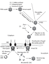
For genome multiplication hepadnaviruses use the transcriptional machinery of the cell that is found within the nucleus. Thus the viral genome has to be transported through the cytoplasm and nuclear pore. The intracytosolic translocation is facilitated by the viral capsid that surrounds the genome and that interacts with cellular microtubules. The subsequent passage through the nuclear pore complexes (NPC) is mediated by the nuclear transport receptors importin alpha and beta. Importin alpha binds to the C-terminus of the capsid protein that comprises a nuclear localization signal (NLS). The exposure of the NLS is regulated and depends upon genome maturation and/or phosphorylation of the capsid protein. As for other karyophilic cargos using this pathway importin alpha interacts with importin beta that facilitates docking of the import complex to the NPC and the passage through the pore. Being a unique strategy, the import of the viral capsid is incomplete in that it becomes arrested inside the nuclear basket, which is a cage-like structure on the karyoplasmic face of the NPC. Presumably only this compartment provides the factors that are required for capsid disassembly and genome release that is restricted to those capsids comprising a mature viral DNA genome.
Figures


References
-
- Janmey PA, Weitz DA. Dealing with mechanics: mechanisms of force transduction in cells. Trends Biochem Sci. 2004;29:364–370. - PubMed
-
- Luby-Phelps K. Physical properties of cytoplasm. Curr Opin Cell Biol. 1994;6:3–9. - PubMed
-
- Zhang X, Clark AF, Yorio T. Heat shock protein 90 is an essential molecular chaperone for nuclear transport of glucocorticoid receptor beta. Invest Ophthalmol Vis Sci. 2006;47:700–708. - PubMed
-
- Giannakakou P, Sackett DL, Ward Y, Webster KR, Blagosklonny MV, Fojo T. p53 is associated with cellular microtubules and is transported to the nucleus by dynein. Nat Cell Biol. 2000;2:709–717. - PubMed
Publication types
MeSH terms
Substances
LinkOut - more resources
Full Text Sources
Research Materials

