Review
. 2007 Jan 7;13(1):65-73.
doi: 10.3748/wjg.v13.i1.65.

Hepatitis B virus morphogenesis

Affiliations
Review

Hepatitis B virus morphogenesis

Volker Bruss. World J Gastroenterol. .

Abstract

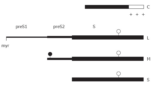
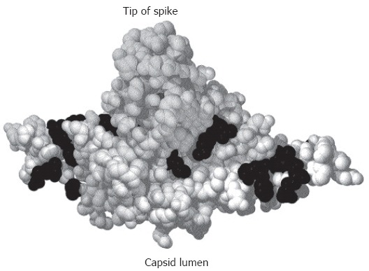
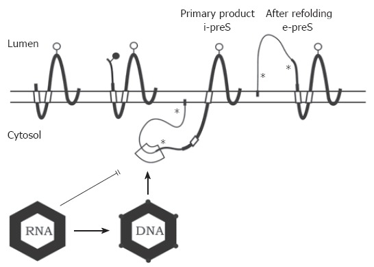
The hepatitis B virus (HBV) particle consists of an envelope containing three related surface proteins and probably lipid and an icosahedral nucleocapsid of approximately 30 nm diameter enclosing the viral DNA genome and DNA polymerase. The capsid is formed in the cytosol of the infected cell during packaging of an RNA pregenome replication complex by multiple copies of a 21-kDa C protein. The capsid gains the ability to bud during synthesis of the viral DNA genome by reverse transcription of the pregenome in the lumen of the particle. The three envelope proteins S, M, and L shape a complex transmembrane fold at the endoplasmic reticulum, and form disulfide-linked homo- and heterodimers. The transmembrane topology of a fraction of the large envelope protein L changes post-translationally, therefore, the N terminal domain of L (preS) finally appears on both sides of the membrane. During budding at an intracellular membrane, a short linear domain in the cytosolic preS region interacts with binding sites on the capsid surface. The virions are subsequently secreted into the blood. In addition, the surface proteins can bud in the absence of capsids and form subviral lipoprotein particles of 20 nm diameter which are also secreted.


Figures

Figure 1
Figure 1
Linear map of the main structural HBV proteins. The C-terminal region (open box) of the capsid protein C is rich in arginine residues (+). The sequence of the small envelope S is also present at the C termini of the M and L protein. The two larger envelope proteins contain the additional N-terminal preS2 and preS2 + preS1 domain, respectively. The L protein is myristylated at glycin 2 (myr), the preS2 domain of M is N-glycosylated (filled circles), and the S domain of all 3 proteins is partially N-glycosylated (open circles).
Figure 2
Figure 2
Crystal structure of a C-terminally truncated C protein dimer[21]. The spike protrudes upwards. The lumen of the capsid would be below the figure. Mutational analysis identified aa (shown in black) where the mutation was compatible with capsid formation and viral DNA synthesis in the lumen of the particle but blocked nucleocapsid envelopment[121].
Figure 3
Figure 3
Transmembrane topology of the HBV envelope proteins and model for envelope-capsid interaction. The transmembrane folding of the S protein is determined by an N-terminal and an internal signal shown as open boxes. The C-terminal domain is hydrophobic and probably embedded in the lipid bilayer (horizontal open bar). The C terminus is oriented towards the ER lumen. The folding of the M protein is similar to S. The preS2 domain of M (thinner line) is located in the ER lumen. In the initial folding of the L protein, the preS domains are located in the cytosol (i-preS). Whether the N-terminal myristate group (filled box) is inserted into the membrane as shown here is unknown. After refolding approximately half of the L chains expose the preS domains at the luminal side of the membrane (e-preS). Open and filled circles: see Figure 1. Asterisks indicate potential but unused N-glycosylation sites in preS of L. A domain in i-preS (boxed area) and in the cytosolic loop of S may interact with the capsid during budding. Immature capsids containing pregenomic RNA are not capable to bud. During viral DNA synthesis, the capsid shell changes (indicated by filled circles at the edges) and becomes competent for envelopment.

References

    1. Acs G, Sells MA, Purcell RH, Price P, Engle R, Shapiro M, Popper H. Hepatitis B virus produced by transfected Hep G2 cells causes hepatitis in chimpanzees. Proc Natl Acad Sci USA. 1987;84:4641–4644. - PMC - PubMed
    1. Yaginuma K, Shirakata Y, Kobayashi M, Koike K. Hepatitis B virus (HBV) particles are produced in a cell culture system by transient expression of transfected HBV DNA. Proc Natl Acad Sci USA. 1987;84:2678–2682. - PMC - PubMed
    1. Gripon P, Rumin S, Urban S, Le Seyec J, Glaise D, Cannie I, Guyomard C, Lucas J, Trepo C, Guguen-Guillouzo C. Infection of a human hepatoma cell line by hepatitis B virus. Proc Natl Acad Sci USA. 2002;99:15655–15660. - PMC - PubMed
    1. Nowak MA, Bonhoeffer S, Hill AM, Boehme R, Thomas HC, McDade H. Viral dynamics in hepatitis B virus infection. Proc Natl Acad Sci USA. 1996;93:4398–4402. - PMC - PubMed
    1. Roingeard P, Sureau C. Ultrastructural analysis of hepatitis B virus in HepG2-transfected cells with special emphasis on subviral filament morphogenesis. Hepatology. 1998;28:1128–1133. - PubMed

MeSH terms