. 2007 Jan 5;3(1):e8.
doi: 10.1371/journal.pgen.0030008. Epub 2006 Nov 30.

A role for Alström syndrome protein, alms1, in kidney ciliogenesis and cellular quiescence

Affiliations

A role for Alström syndrome protein, alms1, in kidney ciliogenesis and cellular quiescence

Guochun Li et al. PLoS Genet. .

Abstract

Premature truncation alleles in the ALMS1 gene are a frequent cause of human Alström syndrome. Alström syndrome is a rare disorder characterized by early obesity and sensory impairment, symptoms shared with other genetic diseases affecting proteins of the primary cilium. ALMS1 localizes to centrosomes and ciliary basal bodies, but truncation mutations in Alms1/ALMS1 do not preclude formation of cilia. Here, we show that in vitro knockdown of Alms1 in mice causes stunted cilia on kidney epithelial cells and prevents these cells from increasing calcium influx in response to mechanical stimuli. The stunted-cilium phenotype can be rescued with a 5' fragment of the Alms1 cDNA, which resembles disease-associated alleles. In a mouse model of Alström syndrome, Alms1 protein can be stably expressed from the mutant allele and is required for cilia formation in primary cells. Aged mice developed specific loss of cilia from the kidney proximal tubules, which is associated with foci of apoptosis or proliferation. As renal failure is a common cause of mortality in Alström syndrome patients, we conclude that this disease should be considered as a further example of the class of renal ciliopathies: wild-type or mutant alleles of the Alström syndrome gene can support normal kidney ciliogenesis in vitro and in vivo, but mutant alleles are associated with age-dependent loss of kidney primary cilia.


Conflict of interest statement

Competing interests. RJG owns stock in Phenomix.

Figures
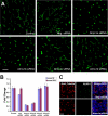
Figure 1
Figure 1. Suppression of Alms1 Expression Alters Primary Cilium Formation in Kidney Epithelial Cells
(A) Elongated cilia, visualized with staining of acetylated tubulin (green), form normally in mIMCD3 cells after mock-transfection, transfection with a negative control siRNA, or transfection with two inactive siRNAs directed against Alms1 (Alms1c and Alms1d). Focal staining of acetylated tubulin without axoneme extension is seen after transfection with two active siRNAs targeting Alms1 (Alms1a and Alms1b). (B) Real-time PCR analysis with two mouse Alms1 probes recognizing the junctions of exons 1 and 2 and exons 12 and 13, respectively: Alms1a and Alms1b siRNAs both cause 70%–80% knockdown of Alms1 mRNA; no effect on Alms1 mRNA was seen with the three siRNAs that were inactive in the ciliogenesis assay. (C) Alms1a siRNA-treated cells lose endogenous Alms1 protein expression. Acet, acetylated. Scale bars, 10 μm.
Figure 2
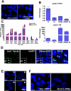
Figure 2. Loss of Alms1 Does Not Affect Transcriptional Changes during Ciliogenesis but Causes Impairment in Flow-Induced Mechanosensation
(A) Confocal microscopic analysis of cilia biogenesis in mIMCD3 cells. Short cilia can be detected at day 3 after transfection. mIMCD3 cells transfected with Alms1a siRNA have stunted cilia at days 3 and 5. Cells were stained with anti-acetylated tubulin (yellow, cilia) and TO-PRO-3 (red, nuclei). (B) Suppression of Alms1 does not affect the upregulation of Bbs4 and Ttc10 during ciliogenesis. N, negative siRNA; 1a, Alms1a siRNA; 1b, Alms1b siRNA. (C) Heat map representation of microarray analysis of mIMCD3 during ciliogenesis. 98 genes with the most dramatic changes in expression showed approximately equivalent expression changes in the presence of a scrambled siRNA control, or in the presence of specific siRNAs that decreased Alms1 mRNA levels and blocked cilia formation. (D) Stunted cilia formed in the presence of Alms1a siRNA (red) lack flow-induced Ca2+ influx in mIMCD3 cells, compared with a negative control siRNA (blue). Representative data are shown for cytosolic calcium change of individual cells in response to mechanical flow. Arrow points to the start of flow.
Figure 3
Figure 3. N-Terminal Alms1 Protein Can Support Cilia Formation
(A) Cotransfection of Alms1a siRNA-treated cells with a 5′ Alms1 cDNA construct rescues primary cilia formation in mIMCD3 cells. (B) Real-time PCR analysis of Alms1a siRNA and N-terminal Alms1-transfected cells. Upper panel: over-expression of the 5′ cDNA does not affect knockdown of endogenous Alms1 mRNA with Alms1a siRNA. Lower panel: knockdown of endogenous Alms1 mRNA does not affect overexpression of the 5′ cDNA. N, negative control siRNA; cDNA, 5′ Alms1 cDNA; 1a, Alms1a siRNA. (C) Stable expression of Alms1 mRNA from the Alms1L2131X/L2131X allele. Real-time PCR analysis of Alms1 gene expression in an Alms1L2131X/L2131X mouse and a wild-type littermate control. (D) The N-terminal mouse Alms1 antibody detects Alms1 mutant protein at the ciliary basal body in primary kidney cells from the Alms1L2131X/L2131X mouse. Shown are low and high magnifications of the ciliated cells. Arrowheads point out the base of cilia. (E) Normal appearance of primary cilia in primary fibroblasts (MEF) and primary kidney cells (PKC) from the Alms1L2131X/L2131X mouse strain. (F) Inhibition of cilia formation in Alms1a siRNA-treated Alms1L2131X/L2131X primary fibroblasts. Scale bars, 10 μm.
Figure 4
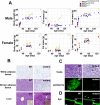
Figure 4. Alms1L2131X/L2131X Mice Recapitulate Human Alström Syndrome
(A) Alms1L2131X/L2131X mice gain more fat mass than heterozygote or wild-type controls but equivalent lean mass. (B) Histological examination of Alms1L2131X/L2131X mice and wild-type littermate control. Insets show oil red O staining of frozen liver sections. (C) Testis H&E sections show degeneration of seminiferous tubules (arrow), which have reduced numbers of germinal cells. Reduced numbers of sperm flagella with decreased length are observed in the epididymus of Alms1L2131X/L2131X mice (anti-acetylated tubulin, green). H&E, hematoxylin-eosin. (D) Rhodopsin staining in the outer nuclear layer cell bodies is seen in rare cells in the Alms1L2131X/L2131X animals (arrows) but not in wild-type littermate controls. Insets illustrate higher magnification images. ONL, outer nuclear layer; IS, inner segment; OS; outer segment. Scale bars, 50 μm.
Figure 5
Figure 5. Kidney Abnormalities in Alms1 Mutant Mice
(A) H&E-stained kidney sections of a 6-mo-old Alms1L2131X/L2131X mouse showing dilated cortex tubules compared with an age-matched wild-type control. Lack of kidney cilia is observed in some tubules in the cortex of Alms1L2131X/L2131X kidney, whereas cilia in the medulla appear normal. H&E, hematoxylin-eosin; Acet, acetylated. (B) Cortex cilia count comparison between Alms1L2131X/L2131X and controls. 300–400 kidney nuclei were examined for each of six fields of Alms1L2131X/L2131X and wild-type controls. The bar chart represents the average and standard deviations of cilia count per 100 kidney cortex epithelial cells from eight mice per group. (C) In the cortex of Alms1L2131X/L2131X kidney, cilia are lost selectively in LTA-labeled tubules but not in aquaporin-2-expressing tubules. (D) Upper panel: clusters of Ki67-positive proliferating epithelial cells in the Alms1L2131X/L2131X kidney, potentially lining the same convoluted tubule. Lower panel: TUNEL staining reveals apoptotic cells in Alms1L2131X/L2131X kidneys but rarely in a wild-type control. Arrow, whole tubule cross sections were labeled by TUNEL, suggesting progression of nephropathy in Alms1L2131X/L2131X mutant kidneys. WT, wild-type. (E) Urinalysis of 3- to 6-mo-old Alms1L2131X/L2131X mice and age-matched littermate controls. Urine from Alms1L2131X/L2131X mice showed slight elevation of protein levels, p = 0.007. Scale bars, 50 μm.

Similar articles

Cited by

References

    1. Marshall JD, Bronson RT, Collin GB, Nordstrom AD, Maffei P, et al. New Alstrom syndrome phenotypes based on the evaluation of 182 cases. Arch Intern Med. 2005;165:675–683. - PubMed
    1. Collin GB, Cyr E, Bronson R, Marshall JD, Gifford EJ, et al. Alms1-disrupted mice recapitulate human Alstrom syndrome. Hum Mol Genet. 2005;14:2323–2333. - PMC - PubMed
    1. Singla V, Reiter JF. The primary cilium as the cell's antenna: Signaling at a sensory organelle. Science. 2006;313:629–633. - PubMed
    1. Davis EE, Brueckner M, Katsanis N. The emerging complexity of the vertebrate cilium: New functional roles for an ancient organelle. Dev Cell. 2006;11:9–19. - PubMed
    1. Wilson PD. Polycystic kidney disease: New understanding in the pathogenesis. Int J Biochem Cell Biol. 2004;36:1868–1873. - PubMed

Publication types

MeSH terms

Associated data