Comparative Study
. 2007 Jan 7;8(1):1.
doi: 10.1186/1465-9921-8-1.

Detection of epithelial to mesenchymal transition in airways of a bleomycin induced pulmonary fibrosis model derived from an alpha-smooth muscle actin-Cre transgenic mouse

Affiliations
Comparative Study

Detection of epithelial to mesenchymal transition in airways of a bleomycin induced pulmonary fibrosis model derived from an alpha-smooth muscle actin-Cre transgenic mouse

Zhuang Wu et al. Respir Res. .

Abstract

Background: Epithelial to mesenchymal transition (EMT) in alveolar epithelial cells (AECs) has been widely observed in patients suffering interstitial pulmonary fibrosis. In vitro studies have also demonstrated that AECs could convert into myofibroblasts following exposure to TGF-beta1. In this study, we examined whether EMT occurs in bleomycin (BLM) induced pulmonary fibrosis, and the involvement of bronchial epithelial cells (BECs) in the EMT. Using an alpha-smooth muscle actin-Cre transgenic mouse (alpha-SMA-Cre/R26R) strain, we labelled myofibroblasts in vivo. We also performed a phenotypic analysis of human BEC lines during TGF-beta1 stimulation in vitro.

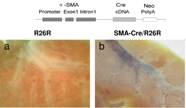
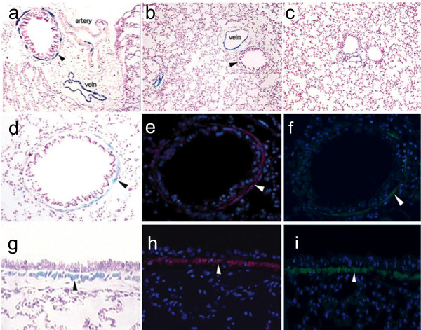
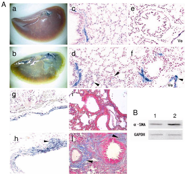
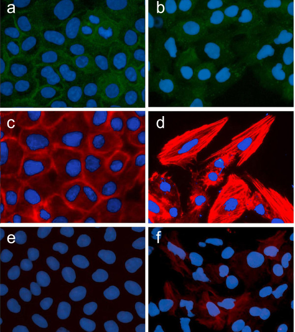
Methods: We generated the alpha-SMA-Cre mouse strain by pronuclear microinjection with a Cre recombinase cDNA driven by the mouse alpha-smooth muscle actin (alpha-SMA) promoter. alpha-SMA-Cre mice were crossed with the Cre-dependent LacZ expressing strain R26R to produce the double transgenic strain alpha-SMA-Cre/R26R. beta-galactosidase (betagal) staining, alpha-SMA and smooth muscle myosin heavy chains immunostaining were carried out simultaneously to confirm the specificity of expression of the transgenic reporter within smooth muscle cells (SMCs) under physiological conditions. BLM-induced peribronchial fibrosis in alpha-SMA-Cre/R26R mice was examined by pulmonary betagal staining and alpha-SMA immunofluorescence staining. To confirm in vivo observations of BECs undergoing EMT, we stimulated human BEC line 16HBE with TGF-beta1 and examined the localization of the myofibroblast markers alpha-SMA and F-actin, and the epithelial marker E-cadherin by immunofluorescence.

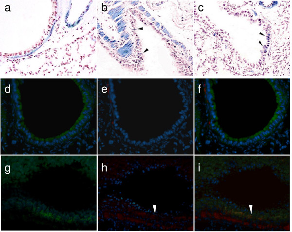
Results: betagal staining in organs of healthy alpha-SMA-Cre/R26R mice corresponded with the distribution of SMCs, as confirmed by alpha-SMA and SM-MHC immunostaining. BLM-treated mice showed significantly enhanced betagal staining in subepithelial areas in bronchi, terminal bronchioles and walls of pulmonary vessels. Some AECs in certain peribronchial areas or even a small subset of BECs were also positively stained, as confirmed by alpha-SMA immunostaining. In vitro, addition of TGF-beta1 to 16HBE cells could also stimulate the expression of alpha-SMA and F-actin, while E-cadherin was decreased, consistent with an EMT.

Conclusion: We observed airway EMT in BLM-induced peribronchial fibrosis mice. BECs, like AECs, have the capacity to undergo EMT and to contribute to mesenchymal expansion in pulmonary fibrosis.


Figures

Figure 1
Figure 1
Transgene construction and βgal staining of lung lobes. A. Transgene fragment for microinjection. The Cre cDNA and a Neo polyadenylylation signal were placed under the control of the mouse α-SMA promoter (-1070 to +2582, including the first exon and part of the first intron). B. Comparison of βgal staining in the bronchi of R26R and α-SMA-Cre/R26R mice. Positive βgal staining (blue color) is observed in the bronchi of α-SMA-Cre/R26R mice (a, 20× magnification), but not in R26R mice (b, 20× magnification).
Figure 2
Figure 2
βgal and immunofluorescent staining in lung tissues of α-SMA-Cre/R26R mice. In βgal stained sections (a, b, c, d, g), intrapulmonary veins were homogeneously stained (a, b) and pulmonary arteries were heterogeneously stained (a). In the main bronchus of pulmonary hilum, unstained ciliated epithelia were surrounded by a βgal stained muscular layer (a, arrowhead), βgal staining was not detected in terminal bronchioles, although small veins were positively stained (c). The thin layer of βgal staining was observed in the sub-epithelial areas of small and medium bronchi, respectively (arrowheads in b, d, g). The βgal stained areas of bronchus (d, g) paralleled the staining pattern for α-SMA (TRITC-labeled, arrowheads in e, h) and SM-MHC (FITC-labeled, arrowheads in f, i) (a-c, 100×, d-i, 400× magnification).
Figure 3
Figure 3
Effects of BLM on lung α-SMA protein levels, ECM deposition and βgal staining. Panel A: βgal and Masson's trichrome-staining in sections of lung tissue shows βgal staining to wholemount left lung lobes of the PBS-treated (a) and the BLM-treated mice (b) (a, b 10× magnification). In moderate bronchi, thickened bronchial wall with homogeneous βgal stained fusiform cells was observed in the BLM-treated lungs (d), and not in the PBS-treated mouse (c), Arrowheads indicate that a few cells in alveolar wall were positively stained (d). In pulmonary bronchioles and vessels, BLM treated lung demonstrated enhanced βgal expression (f, h), compared with that of PBS treated lung (e, g). βgal stained venous wall was thickened (arrowhead in f) and the positively stained cells infiltrated outwards (arrowhead in h). In the Masson's trichrome-stained lung sections (i, j), extensive collagen staining (Blue color) was seen in BLM treated lung (arrowheads in j), but not in the control with PBS (i). (c-j 400× magnification). Panel B: Western blot analysis of protein extracts from lower right lobes of the BLM treated mice (2) and control mice (1).
Figure 4
Figure 4
βgal and αSMA positively stained bronchial epithelial cells in the α-SMA-Cre R26R mice treated with BLM. βgal stained lung sections of α-SMA-Cre/R26R mice without and with BLM treatment (a-c). The section from BLM treated mice showed a few βgal stained bronchial epithelial cells (arrowheads in b and c), but not from PBS treated mice (a). Double immunofluorescent staining for α-SMA and E-Cadherin was performed on the sections from PBS (d-f) or BLM (g-i) treated mice. d, g: FITC-labeled E-cadherin; e, h: TRITC labeled α-SMA. Positive double immunofluorescent staining (g, h, i) was observed in the bronchial epithelial cells lining the bronchioles of the BLM-treated lung where βgal staining was detected as above, but not in the control (d, e, f). The images of (d) and (e), or (g) and (h) were merged into (f) and (i). The red fluorescence (h arrowhead) indicated the positive α-SMA staining and yellow fluorescent staining (i arrowhead) indicated that the epithelial cells positively co-stained with α-SMA and E-Cadherin. (all images are 400× magnification).
Figure 5
Figure 5
Phenotypic analysis of the human bronchial epithelial cell line (16HBE) following exposure to TGF-β1. Immunofluorescent staining for E-cadherin (a, b) showed that exposure to TGF-β1 (b) resulted in an apparent reduction and redistribution of E-cadherin from intercellular junction areas into cytoplasm, compared to control (a). Mesenchymal marker F-actin, was faintly stained at the cell margin in the control (c), whereas the staining was substantially enhanced and abundantly located throughout cytoplasm after TGF-β1 stimulation (d). Immunofluorescent staining for (-SMA was not detected in the cells under basal conditions (e), but was observable in a few cells after TGF-β1 exposure (f).

Similar articles

Cited by

References

    1. Kuhn C, McDonald JA. The roles of the myofibroblast in idiopathic pulmonary fibrosis. Ultrastructural and immunohistochemical features of sites of active extracellular matrix synthesis. Am J Pathol. 1991;138(5):1257–1265. - PMC - PubMed
    1. Schissel SL, Layne MD. Telomerase, myofibroblasts, and pulmonary fibrosis. Am J Respir Cell Mol Biol. 2006;34(5):520–522. doi: 10.1165/rcmb.F312. - DOI - PubMed
    1. White ES, Atrasz RG, Hu B, Phan SH, Stambolic V, Mak TW, Hogaboam CM, Flaherty KR, Martinez FJ, Kontos CD, et al. Negative regulation of myofibroblast differentiation by PTEN (Phosphatase and Tensin Homolog Deleted on chromosome 10) Am J Respir Crit Care Med. 2006;173(1):112–121. doi: 10.1164/rccm.200507-1058OC. - DOI - PMC - PubMed
    1. Chambers RC, Leoni P, Kaminski N, Laurent GJ, Heller RA. Global expression profiling of fibroblast responses to transforming growth factor-betal reveals the induction of inhibitor of differentiation-1 and provides evidence of smooth muscle cell phenotypic switching. Am J Pathol. 2003;162(2):533–546. - PMC - PubMed
    1. Oda D, Gown AM, Vande Berg JS, Stern R. The fibroblast-like nature of myofibroblasts. Exp Mol Pathol. 1988;49(3):316–329. doi: 10.1016/0014-4800(88)90004-4. - DOI - PubMed

Publication types

MeSH terms