

Further investigation of the mechanism of Doxorubicin release from P105 micelles using kinetic models
- PMID: 17207611
- PMCID: PMC2262855
- DOI: 10.1016/j.colsurfb.2006.11.006
Further investigation of the mechanism of Doxorubicin release from P105 micelles using kinetic models
Abstract
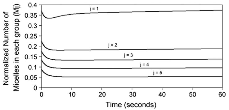
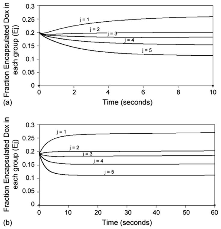
The kinetics of the release of Doxorubicin from Pluronic P105 micelles during ultrasonication and its subsequent re-encapsulation upon cessation of insonation were investigated. Four mechanisms are proposed to explain the acoustically-triggered Doxorubicin (Dox) release and re-encapsulation from Pluronic P105 micelles. The four mechanisms are: micelle destruction; destruction of cavitating nuclei; reassembly of micelles, and the re-encapsulation of Dox. The first mechanism, the destruction of micelles during insonation, causes the release of Dox into solution. The micelles are destroyed because of cavitation events produced by collapsing nuclei, or bubbles in the insonated solution. The second mechanism, the slow destruction of cavitating nuclei, results in a slow partial recovery phase, when a small amount of Dox is re-encapsulated. The third and fourth mechanisms, the reassembly of micelles and the re-encapsulatin of Dox, are independent of ultrasound. These two mechanism are responsible for maintaining the drug release at a partial level, and for recovery after insonation ceases. A normal distribution was used to describe micellar size. Parameters for the model were determined based upon the best observed fit to experimental data. The resulting model provides a good approximation to experimental data for the release of Dox from Pluronic P105 micelles.
Figures


Similar articles
-
Kinetics and Thermodynamics of Acoustic Release of Doxorubicin from Non-stabilized polymeric Micelles.Colloids Surf A Physicochem Eng Asp. 2010 Apr 20;359(1-3):18-24. doi: 10.1016/j.colsurfa.2010.01.044. Colloids Surf A Physicochem Eng Asp. 2010. PMID: 20495608 Free PMC article.
-
The use of ultrasound and micelles in cancer treatment.J Nanosci Nanotechnol. 2008 May;8(5):2205-15. doi: 10.1166/jnn.2008.225. J Nanosci Nanotechnol. 2008. PMID: 18572632 Free PMC article. Review.
-
Ultrasonic release of doxorubicin from Pluronic P105 micelles stabilized with an interpenetrating network of N,N-diethylacrylamide.J Control Release. 2002 Oct 4;83(2):303-5. doi: 10.1016/s0168-3659(02)00203-1. J Control Release. 2002. PMID: 12363455
-
Sequestration and ultrasound-induced release of doxorubicin from stabilized Pluronic P105 micelles.Drug Deliv. 2002 Oct-Dec;9(4):253-8. doi: 10.1080/10717540260397873. Drug Deliv. 2002. PMID: 12511204
-
Modeling and sensitivity analysis of acoustic release of Doxorubicin from unstabilized pluronic P105 using an artificial neural network model.Technol Cancer Res Treat. 2007 Feb;6(1):49-56. doi: 10.1177/153303460700600107. Technol Cancer Res Treat. 2007. PMID: 17241100 Free PMC article.
Cited by
-
Kinetics and Thermodynamics of Acoustic Release of Doxorubicin from Non-stabilized polymeric Micelles.Colloids Surf A Physicochem Eng Asp. 2010 Apr 20;359(1-3):18-24. doi: 10.1016/j.colsurfa.2010.01.044. Colloids Surf A Physicochem Eng Asp. 2010. PMID: 20495608 Free PMC article.
-
Degradation kinetics of stabilized Pluronic micelles under the action of ultrasound.J Control Release. 2009 Aug 19;138(1):45-8. doi: 10.1016/j.jconrel.2009.04.018. Epub 2009 Apr 21. J Control Release. 2009. PMID: 19389432 Free PMC article.
-
Ultrasonic-activated micellar drug delivery for cancer treatment.J Pharm Sci. 2009 Mar;98(3):795-811. doi: 10.1002/jps.21444. J Pharm Sci. 2009. PMID: 18506804 Free PMC article. Review.
-
The use of ultrasound and micelles in cancer treatment.J Nanosci Nanotechnol. 2008 May;8(5):2205-15. doi: 10.1166/jnn.2008.225. J Nanosci Nanotechnol. 2008. PMID: 18572632 Free PMC article. Review.
-
Synthesis and characterization of arginine-glycine-aspartic peptides conjugated poly(lactic acid-co-L-lysine) diblock copolymer.J Mater Sci Mater Med. 2008 Mar;19(3):1275-81. doi: 10.1007/s10856-007-3237-9. Epub 2007 Aug 20. J Mater Sci Mater Med. 2008. PMID: 17705054
References
-
- Rapoport N, Caldwell K. Structural transitions in micellar solutions of Pluronic P-105 and their effect on the conformation of dissolved Cytochrome C: an electron paramagnetic resonance investigation. Colloid Surf B: Biointerfaces. 1994;3:217–228.
-
- Husseini GA, Christensen DA, Rapoport NY, Pitt WG. Ultrasonic release of doxorubicin from Pluronic P105 micelles stabilized with an interpenetrating network of N,N-diethylacrylamide. J Control Release. 2002;83(2):302–304. - PubMed
-
- Husseini GA, Myrup GD, Pitt WG, Christensen DA, Rapoport NY. Factors affecting acoustically-triggered release of drugs from polymeric micelles. J Control Release. 2000;69:43–52. - PubMed
-
- Husseini GA, et al. DNA damage induced by micellar-delivered doxoru-bicin and ultrasound: comet assay study. Cancer Lett. 2000;154:211–216. - PubMed
MeSH terms
Substances
Grants and funding
LinkOut - more resources
Full Text Sources
