. 2007 Apr;46(4):293-301.
doi: 10.1016/j.vph.2006.11.003. Epub 2006 Nov 18.

Evidence supporting changes in Nogo-B levels as a marker of neointimal expansion but not adaptive arterial remodeling

Affiliations

Evidence supporting changes in Nogo-B levels as a marker of neointimal expansion but not adaptive arterial remodeling

Jacek J Paszkowiak et al. Vascul Pharmacol. 2007 Apr.

Abstract

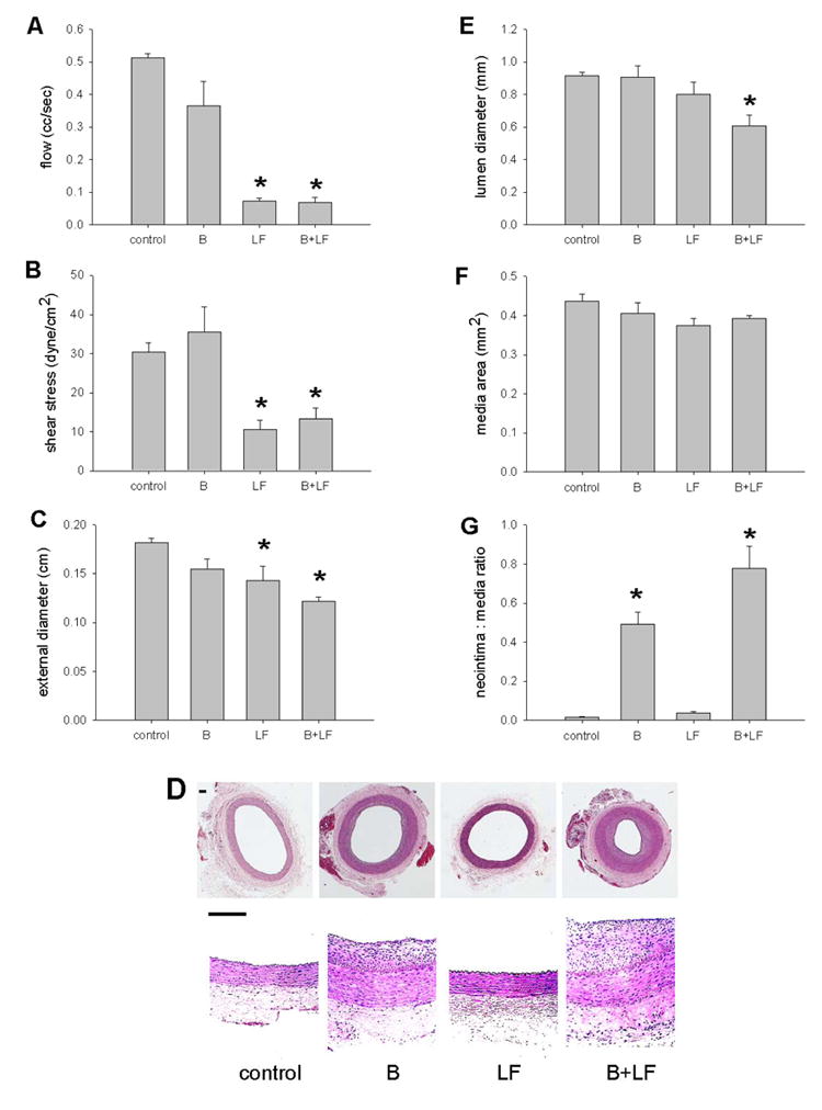
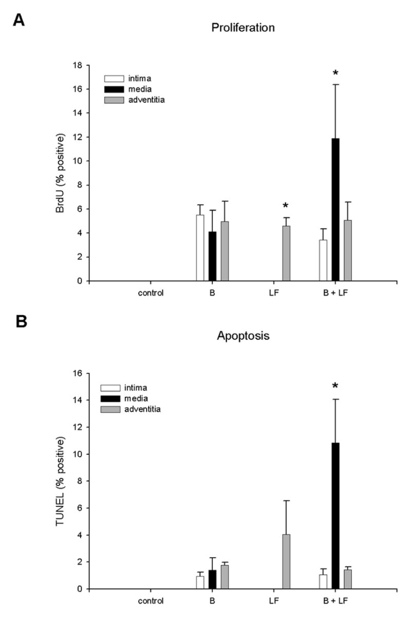
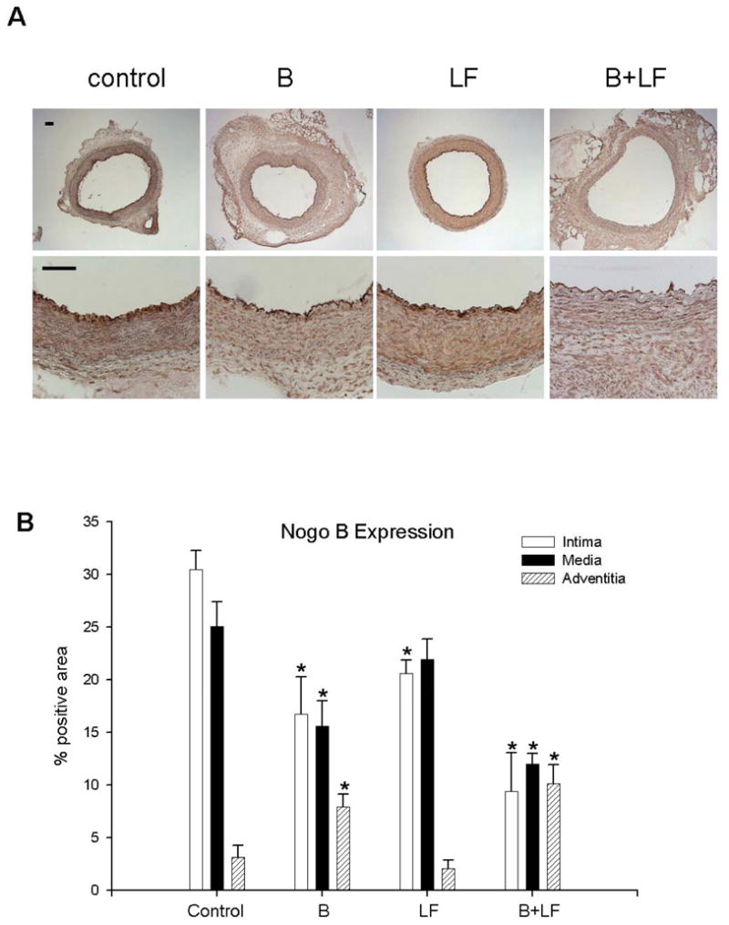
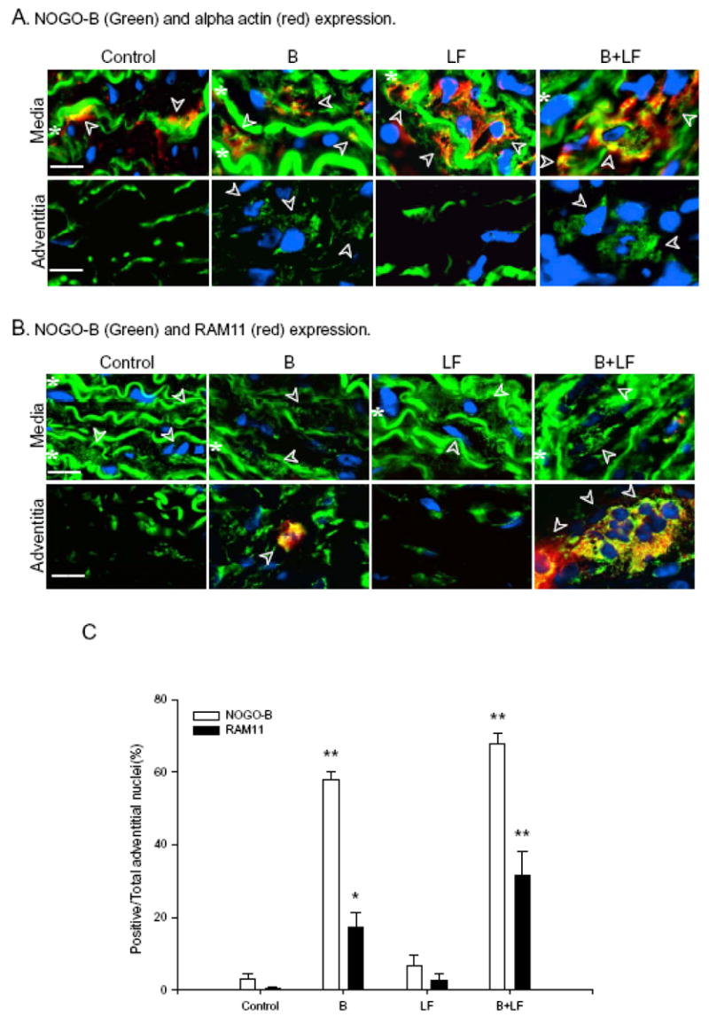
Both neointimal hyperplasia and inward remodeling contribute to restenosis and lumen loss. Nogo-B has been recently described as an inhibitor of vascular injury and neointimal hyperplasia. To determine whether Nogo-B expression may be a mediator of inward remodeling, we examine the localization of expression of Nogo-B in an in vivo model that examines both neointimal hyperplasia and inward remodeling. The rabbit carotid artery was subjected to balloon injury, outflow branch ligation to reduce flow, or both balloon injury and reduction in flow. In balloon injury-induced neointimal hyperplasia Nogo-B expression was reduced in the intima and media but stimulated in the adventitia. In low flow-induced inward remodeling medial Nogo-B expression was not reduced and adventitial Nogo-B expression was not stimulated. Low flow significantly augmented balloon injury-induced neointimal hyperplasia and was accompanied by reduced intimal and medial Nogo-B expression, and increased adventitial Nogo-B expression in both smooth muscle cells and macrophages. Low flow-induced inward remodeling is not associated with changes in medial Nogo-B expression and is distinct from injury-induced neointimal hyperplasia. Pharmacological strategies to inhibit neointimal hyperplasia and restenosis using normal flow models may only partially account for lumen loss and therefore may not accurately predict responses in patients with extensive outflow disease.


Figures

Figure 1
Figure 1
Effects of interactions on vessel size and hemodynamics (21d). A) Reduced flow in animals treated with low flow (LF) (*, p<.0001, post-hoc) or balloon injury and low flow (B + LF) (*, p<.0001) compared to control animals or animals treated with balloon injury (B) alone (p<.0001, ANOVA; n=21). B) Reduced shear stress in animals treated with LF (*, p=.01, post-hoc) or B+LF (*, p=.03) compared to control animals or animals treated with B alone (p=.001, ANOVA). C) Reduced diameter in animals treated with LF (*, p=.04, post-hoc) or B+LF (*, p=.003) compared to control animals or animals treated with B alone (p=.02, ANOVA). The 21% decrease in diameter in the LF group is similar to previous reports.(Langille and O'Donnell 1986) D) Representative photomicrographs of low and high power magnification of vessels (control, B, LF, B+LF). Scale bar, 100 μm. E) Reduced lumen diameter in animals treated with B+LF (*, p=.02, post-hoc) compared to control animals (p=.02, ANOVA). F) Similar media area between animal groups (p=.28, ANOVA). G) Increased neointima:media ratio in animals treated with B or B+LF (p<.0001, ANOVA). The increased neointima:media ratio in B+LF, compared to the increase in B, is significant (*, p=.01, post-hoc).
Figure 2
Figure 2
Proliferation and apoptosis in vessels (7d). A) The percentage of cells staining positively for BrdU is plotted according to location within the vessel wall. The difference between the adventitia and the other layers in the LF group is significant (*; p=.005, post-hoc); the difference between the media in the B+LF group and the media of the other groups is significant (*; p=.02, post-hoc; n=12). B) The percentage of cells staining positively for TUNEL is plotted according to location within the vessel wall. The difference between the media in the B+LF group and the media of the other groups is significant (*; p=.01, post-hoc).
Figure 3
Figure 3
Nogo-B expression in the rabbit carotid artery (7d). A) Representative photomicrographs demonstrating immunohistochemistry for Nogo-B in the rabbit carotid artery (day 7), treated with: control, B, LF, B+LF. Scale bar, 100 μm. B) Quantitative analysis of Nogo-B density in the intima, media and adventitia.
Figure 4
Figure 4
Nogo-B localization in the rabbit carotid artery (7d). A) Representative photomicrographs demonstrating immunofluorescence for Nogo-B (green), alpha-actin (red), or both merged (yellow) in the rabbit carotid artery (day 7), treated with: control, B, LF, B+LF. Nuclei are indicated by DAPI (blue) fluorescence. Row I: Media; Row II: Adventitia. Scale bar, 20 μm. Arrowheads represent cells staining for Nogo-B and alpha-actin (media), and cells staining for Nogo-B but not alpha-actin (adventitia); white stars represent autofluorescence of the medial elastic laminae. B) Representative photomicrographs demonstrating merged immunofluorescence for Nogo-B (green), RAM-11 (red), or both merged (yellow); and nuclei with DAPI (blue). Row I: Media; Row II: Adventitia. Scale bar, 20 μm. Arrowheads represent cells staining for Nogo-B but not RAM-11 (media), and cells staining for both Nogo-B and RAM-11 (adventitia); white stars represent autofluorescence of the medial elastic laminae. C) Bar graph demonstrating the percentage of cells staining positively for Nogo-B (white bars) and RAM-11 (black bars) in the adventitia of rabbits treated with: control, B, LF, B+LF.

References

    1. Acevedo L, Yu J, et al. A new role for Nogo as a regulator of vascular remodeling. Nat Med. 2004;10(4):382–8. - PubMed
    1. Austin GE, Ratliff NB, et al. Intimal proliferation of smooth muscle cells as an explanation for recurrent coronary artery stenosis after percutaneous transluminal coronary angioplasty. J Am Coll Cardiol. 1985;6(2):369–75. - PubMed
    1. Bauters C, Isner JM. The biology of restenosis. Prog Cardiovasc Dis. 1997;40(2):107–16. - PubMed
    1. Booth RF, Martin JF, et al. Rapid development of atherosclerotic lesions in the rabbit carotid artery induced by perivascular manipulation. Atherosclerosis. 1989;76(2–3):257–68. - PubMed
    1. Cho A, Mitchell L, et al. Effects of changes in blood flow rate on cell death and cell proliferation in carotid arteries of immature rabbits. Circ Res. 1997;81(3):328–37. - PubMed

Publication types

MeSH terms