Hybrid homology modeling and mutational analysis of cytochrome P450C24A1 (CYP24A1) of the Vitamin D pathway: insights into substrate specificity and membrane bound structure-function
- PMID: 17207766
- PMCID: PMC1978416
- DOI: 10.1016/j.abb.2006.11.018
Hybrid homology modeling and mutational analysis of cytochrome P450C24A1 (CYP24A1) of the Vitamin D pathway: insights into substrate specificity and membrane bound structure-function
Abstract
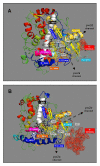
Cytochrome P450C24A1 (CYP24A1), a peripheral inner mitochondrial membrane hemoprotein and candidate oncogene, regulates the side-chain metabolism and biological function of vitamin D and many of its related analog drugs. Rational mutational analysis of rat CYP24A1 based on hybrid (2C5/BM-3) homology modeling and affinity labeling studies clarified the role of key domains (N-terminus, A', A, and F-helices, beta3a strand, and beta5 hairpin) in substrate binding and catalysis. The scope of our study was limited by an inability to purify stable mutant enzyme targeting soluble domains (B', G, and I-helices) and suggested greater conformational flexibility among CYP24A1's membrane-associated domains. The most notable mutants developed by modeling were V391T and I500A, which displayed defective-binding function and profound metabolic defects for 25-hydroxylated vitamin D3 substrates similar to a non-functional F-helix mutant (F249T) that we previously reported. Val-391 (beta3a strand) and Ile-500 (beta5 hairpin) are modeled to interact with Phe-249 (F-helix) in a hydrophobic cluster that directs substrate-binding events through interactions with the vitamin D cis-triene moiety. Prior affinity labeling studies identified an amino-terminal residue (Ser-57) as a putative active-site residue that interacts with the 3beta-OH group of the vitamin D A-ring. Studies with 3-epi and 3-deoxy-1,25(OH)2D3 analogs confirmed interactions between the 3beta-OH group and Ser-57 effect substrate recognition and trafficking while establishing that the trans conformation of A-ring hydroxyl groups (1alpha and 3beta) is obligate for high-affinity binding to rat CYP24A1. Our work suggests that CYP24A1's amphipathic nature allows for monotopic membrane insertion, whereby a pw2d-like substrate access channel is formed to shuttle secosteroid substrate from the membrane to the active-site. We hypothesize that CYP24A1 has evolved a unique amino-terminal membrane-binding motif that contributes to substrate specificity and docking through coordinated interactions with the vitamin D A-ring.
Figures


Similar articles
-
Generation of a homology model for the human cytochrome P450, CYP24A1, and the testing of putative substrate binding residues by site-directed mutagenesis and enzyme activity studies.Arch Biochem Biophys. 2007 Apr 15;460(2):177-91. doi: 10.1016/j.abb.2006.11.030. Epub 2006 Dec 13. Arch Biochem Biophys. 2007. PMID: 17224124
-
Crystal structure of CYP24A1, a mitochondrial cytochrome P450 involved in vitamin D metabolism.J Mol Biol. 2010 Feb 19;396(2):441-51. doi: 10.1016/j.jmb.2009.11.057. Epub 2009 Dec 1. J Mol Biol. 2010. PMID: 19961857 Free PMC article.
-
Rat cytochrome P450C24 (CYP24A1) and the role of F249 in substrate binding and catalytic activity.Arch Biochem Biophys. 2004 May 15;425(2):133-46. doi: 10.1016/j.abb.2004.01.025. Arch Biochem Biophys. 2004. PMID: 15111121
-
[Recent studies on vitamin D metabolizing enzymes].Clin Calcium. 2006 Jul;16(7):1129-35. Clin Calcium. 2006. PMID: 16816472 Review. Japanese.
-
25-Hydroxyvitamin D-24-hydroxylase (CYP24A1): its important role in the degradation of vitamin D.Arch Biochem Biophys. 2012 Jul 1;523(1):9-18. doi: 10.1016/j.abb.2011.11.003. Epub 2011 Nov 12. Arch Biochem Biophys. 2012. PMID: 22100522 Review.
Cited by
-
1,25-(OH)2D-24 Hydroxylase (CYP24A1) Deficiency as a Cause of Nephrolithiasis.Clin J Am Soc Nephrol. 2013 Apr;8(4):649-57. doi: 10.2215/CJN.05360512. Epub 2013 Jan 4. Clin J Am Soc Nephrol. 2013. PMID: 23293122 Free PMC article.
-
Cytochrome P450-mediated metabolism of vitamin D.J Lipid Res. 2014 Jan;55(1):13-31. doi: 10.1194/jlr.R031534. Epub 2013 Apr 6. J Lipid Res. 2014. PMID: 23564710 Free PMC article. Review.
-
Regulation of zebrafish fin regeneration by vitamin D signaling.Dev Dyn. 2021 Sep;250(9):1330-1339. doi: 10.1002/dvdy.261. Epub 2020 Oct 26. Dev Dyn. 2021. PMID: 33064344 Free PMC article.
-
Intestinal Cyp24a1 regulates vitamin D locally independent of systemic regulation by renal Cyp24a1 in mice.J Clin Invest. 2024 Dec 17;135(4):e179882. doi: 10.1172/JCI179882. J Clin Invest. 2024. PMID: 39688907 Free PMC article.
-
Association of the CYP24A1-rs2296241 polymorphism of the vitamin D catabolism enzyme with hormone-related cancer risk: a meta-analysis.Onco Targets Ther. 2015 May 22;8:1175-83. doi: 10.2147/OTT.S80311. eCollection 2015. Onco Targets Ther. 2015. PMID: 26045671 Free PMC article. Review.
References
-
- Sakaki T, Kagawa N, Yamamoto K, Inouye K. Metabolism of vitamin D3 by cytochromes P450. Front Biosci. 2005;10:119–34. - PubMed
-
- Prosser DE, Jones G. Enzymes involved in the activation and inactivation of vitamin D. Trends Biochem Sci. 2004;12:664–73. - PubMed
-
- Yano JK, Hsu MH, Griffin KJ, Stout CD, Johnson EF. Structures of human microsomal cytochrome P450 2A6 complexed with coumarin and methoxsalen. Nat Struct Mol Biol. 2005;12:822–23. - PubMed
-
- Schoch GA, Yano JK, Wester MR, Griffin KJ, Stout CD, Johnson EF. Structure of human microsomal cytochrome P450 2C8. Evidence for a peripheral fatty acid binding site. J Biol Chem. 2003;279:9497–503. - PubMed
-
- Wester MR, Yano JK, Schoch GA, Yang C, Griffin KJ, Stout CD, Johnson EF. The structure of human cytochrome P450 2C9 complexed with flurbiprofen at 2.0-A resolution. J Biol Chem. 2004;279:35630–7. - PubMed
Publication types
MeSH terms
Substances
Grants and funding
LinkOut - more resources
Full Text Sources
Medical

