. 2007 Jun;82(3):234-44.
doi: 10.1016/j.yexmp.2006.10.007. Epub 2007 Jan 4.

Loss of nuclear p21(Cip1/WAF1) during neoplastic progression to metastasis in gamma-irradiated p21 hemizygous mice

Affiliations

Loss of nuclear p21(Cip1/WAF1) during neoplastic progression to metastasis in gamma-irradiated p21 hemizygous mice

Robert W Engelman et al. Exp Mol Pathol. 2007 Jun.

Abstract

p21(Cip1/WAF1) localizes to the nucleus in response to gamma-irradiation induced DNA damage and mediates a G(1) checkpoint arrest. Although gamma-irradiated p21(+/-) mice develop a broad spectrum of tumors, gamma-irradiated p21(-/-) mice develop significantly more metastatic cancers. To evaluate the expression of p21 in tissues prone or resistant to tumorigenesis as a function of gamma-irradiation, and to determine whether phenotypic loss of p21 heterozygosity occurs during tumor progression in p21(+/-) mice, tissues and tumors from gamma-irradiated mice were evaluated immunohistochemically. The percentage of tumors in p21(+/-) mice that were nuclear p21-positive declined with progression to metastasis (p<0.0001). Benign tumors were more often p21-positive and comprised of larger subsets of nuclear p21-positive cells than were malignant tumors of the same histopathological type, while metastatic cancers were nuclear p21-negative (p=0.0003). Even when a primary cancer was comprised of a subset of nuclear p21-positive cells, the metastatic foci of that same cancer were nuclear p21-negative. Mesenchymal tumors, though rare, were more likely metastatic than were epithelial tumors (p=0.0004), and these were invariably nuclear p21-negative. Prepubescent epithelial tissues from which most tumors later originated in mice with reduced p21 gene dosage (i.e., harderian gland, ovary, small intestine, and lung) were p21 expressive within 4 h of gamma-irradiation (p=0.0625), so that p21/Ki67 ratios increased post-gamma-irradiation (p=0.03). In contrast, p21 did not localize to nuclei of cortical thymocytes, a tissue where tumorigenesis was not augmented by reduced p21 gene dosage. Cellular subclones of malignant tumors, especially those of mesenchymal cell origin, which lack nuclear p21 may more readily acquire the genetic alterations of the metastatic phenotype.


Figures

Figure 1
Figure 1
A broad spectrum of metastatic cancers developed in γ-irradiated mice only in the absence of p21 expression, either in p21-/- mice or p21-negative cancer metastases of p21+/- mice. Most metastatic cancers were mesenchymal in origin. Epithelial cancers that were metastatic included an (A) adrenal pheochromocytoma metastatic to the lung, a (B) harderian gland adenocarcinoma metastatic to the lung, a (C) hepatoblastoma with multiple intra-hepatic metastases, a (D) mucicarmine-negative, faintly keratin-positive undifferentiated carcinoma metastatic to the liver from a mouse with a small intestinal adenocarcinoma as the only detectable primary cancer, and a (E) malignant granulosa cell tumor metastatic to the lung. More metastatic mesenchymal cancers developed in γ-irradiated p21+/- and p21-/- mice, including a (F) leiomyosarcoma of undetermined primary site metastatic to the lymph node, an (G) osteogenic sarcoma of undetermined primary site metastatic to the liver, several (H) thymic malignant lymphoma here infiltrating the lung, a (I) hemangiosarcoma metastatic to the lung, a (J) histiocytic sarcoma metastatic to the pancreas, and an (K) anaplastic spindle cell tumor that effaced normal hepatic parenchyma, surrounded a remnant cord of hepatocytes (arrows), and exhibited smooth muscle differentiation (upper left), and in other areas of the same cancer appeared as a well-differentiated (L) leiomyosarcoma metastatic to the liver. Mouse genotype is indicated above each tumor.
Figure 2
Figure 2
Nuclear p21 expression levels and patterns in individual primary tumors. Clusters of p21-positive cells were present along advancing margins of (A) bronchioloalveolar carcinoma, and near the lumenal surface of (B) small intestinal adenocarcinoma, or were present diffusely throughout a (C) harderian gland adenocarcinoma at moderate levels, or a (D) malignant granulosa cell tumor at minimal levels.
Figure 3
Figure 3
Subjacent sections of primary tumors immunostained for p21 or Ki-67. A comparable subset of tumor cells of a harderian gland adenoma immunostained for (A) nuclear p21 or (B) Ki-67, but cells of the adjacent normal glandular parenchyma (left edge of each A & B) rarely stained for either marker. In contrast, (C) a clustered subset of tumor cells near the lumenal surface (arrow) of a small intestinal adenocarcinoma immunostained for nuclear p21, but most tumor cells did not, and only rare cells of the adjacent normal mucosa did (rectangle in upper right), while (D) most tumor cells and numerous cells of the adjacent normal mucosal crypts and lower villi stained for Ki-67.
Figure 4
Figure 4
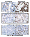
Subjacent sections of metastatic cancers immunostained for p21 (A, C, E) or Ki-67 (B, D, F). Malignant lymphoma (A), here shown infiltrating the kidney, were negative for nuclear p21, although an occasional renal tubular cell nucleus (arrow) was nuclear p21-positive, and (B) most malignant lymphoma cancer cells were Ki67-positive. Cancer cells of all nineteen pulmonary metastatic foci of a metastatic osteogenic sarcoma (C) were negative for nuclear p21, although (D) a moderate subpopulation of osteogenic sarcoma cancer cells were Ki67-positive. Fewer than 3% of cancer cells of a lymph node metastatic focus of a leiomyosarcoma (E) stained faintly for nuclear p21, but these same cancer cells (F) were moderately Ki67-positive.
Figure 5
Figure 5
Nuclear p21 and Ki67 immunostaining levels and patterns of an adrenal gland pheochromocytoma (A-C) that was metastatic to the lung (D-F) and liver (G-I). Metastatic pheochromocytoma cancer cells (arrows) compressed the adjacent normal pulmonary (D) and hepatic (G) parenchyma. Clusters of primary tumor cells were moderately p21-positive (B) and Ki67-positive (C). In contrast, metastatic cancer cells were negative for nuclear p21 (E & H), but moderately Ki67-positive in pulmonary metastatic foci (F), or rarely Ki67-positive (I, arrows) in hepatic metastatic foci.
Figure 6
Figure 6
Nuclear p21 prior to (A, D, G, J) and 4 hours after (B, E, H, K) γ-irradiation, and Ki67 (C, F, I, L) expression 4 hours after γ-irradiation in prepubescent mice. Prior to γ-irradiation, nuclear p21 was only localized in limited tissues, including minimal cells of the oviduct (D, arrow) or exocrine pancreas (G, lower arrow), or in rare cells of the small intestinal crypts (G, upper arrow), but was not present in the cortical thymus (J, left of diagonal). Epithelial tissues prone to tumorigenesis with reduced p21 gene dosage were comprised of a moderate subset of nuclear p21-positive cells after γ-irradiation (B, E, H), but the cortical thymus was not (K, left of diagonal). Nuclear p21 was localized in (B) epithelial cells of the harderian gland ducts and alveoli, in (E) follicular granulosa and theca cells of the ovary, and in (H) mucosal epithelial cells of the upper crypt and lower villus of the small intestine, as well as acinar cells of the exocrine pancreas (H, lower left), and in rare, scattered, heterogeneous cells of the thymic medulla (K, right of diagonal), but not in cortical thymocytes (K, left of diagonal) after γ-irradiation, where numerous apoptotic cells and bodies were evident. Each tissue had moderate to marked pools of replicating Ki67-positive cells (C, F, I, L).

Similar articles

Cited by

References

    1. Pines J. Cyclins and Cyclin-Dependent Kinases: A Biochemical View. Biochem J. 1995;308:697–711. - PMC - PubMed
    1. Morgan DO. Principles of CDK Regulation. Nature. 1995;374:131–134. - PubMed
    1. Sherr CJ, Roberts JM. CDK inhibitors: positive and negative regulators of G1-phase progression. Genes Dev. 1999;13:1501–1512. - PubMed
    1. El-Deiry WS, Tokino T, Velculescu VE, Levy DB, Parsons R, Trent JM, Lin D, Mercer WE, Kinzler KW, Vogelstein B. WAF1, a potential mediator of p53 tumor suppression. Cell. 1993;75:817–825. - PubMed
    1. Brugarolas J, Chandrasekaran C, Gordon JI, Beach D, Jacks T, Hannon GJ. Radiation-induced cell cycle arrest compromised by p21 deficiency. Nature. 1995;377:552–557. - PubMed

Publication types

MeSH terms

Substances

LinkOut - more resources