

Review
doi: 10.1016/j.tibs.2006.12.006.
Epub 2007 Jan 5.
ATP synthase--the structure of the stator stalk
Affiliations
- PMID: 17208001
- PMCID: PMC2570231
- DOI: 10.1016/j.tibs.2006.12.006
Item in Clipboard
Review
ATP synthase--the structure of the stator stalk
Trends Biochem Sci.
2007 Feb.
Abstract
ATP synthase synthesizes ATP from ADP and inorganic phosphate using a unique rotary mechanism whereby two subcomplexes move relative to each other, powered by a proton or sodium gradient. The non-rotating parts of the machinery are held together by the "stator stalk". The recent resolution of the structure of a major portion of the stator stalk of mitochondrial ATP synthase represents an important step towards a structural model for the ATP synthase holoenzyme.
Figures
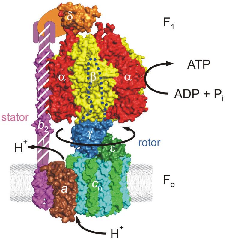
The rotary mechanism of ATP synthase. A model of the E. coli enzyme is shown (taken from Ref. [14], with permission). The portions of the enzyme for which a high-resolution structural model was available are shown in surface representation. The individual structural models (α3β3γε [23], N-terminal domain of δ bound to N-terminus of α [13], acn [24], membrane portion of b [8], “dimerization domain” of b [9]) were docked by eye. Structural data for parts of a and b as well as the C-terminal domain of δ were lacking. ATP synthase can be separated into a membrane-standing subcomplex, Fo, which, in E. coli, consists of subunits ab2cn (brown, pink, and alternating cyan/light green, respectively) and a soluble subcomplex, F1, consisting of subunits α3β3γδε (red, yellow, blue, orange, and green, respectively). A newer, more mechanically-based division differentiates between the “rotor” (in E. coli, subunits γεcn) and the “stator” (subunits α3β3δab2). The dashed blue line indicates the part of γ hidden from view in the center of the α3β3 ring. Flow of protons, down a gradient, through channels in the a subunit at the interface to the cn ring drives the rotor, whose movement results in synthesis of ATP from ADP and Pi in the catalytic nucleotide binding sites on the β subunits and release of product ATP from these sites. The enzyme can also run in reverse, hydrolyzing ATP to generate a proton gradient. In the mitochondrial enzyme, the b dimer is replaced by a single b subunit plus subunits d and F6. The b/d/F6 subcomplex whose structure was resolved by Kane Dickson et al. [10] corresponds to the hatched area. The mitochondrial counterparts of E. coli subunits δ and ε are termed OSCP (“oligomycin-sensitivity conferring protein”) and δ, respectively. Figures 1 and 2A were created with PyMOL (pymol.sourceforge.net).
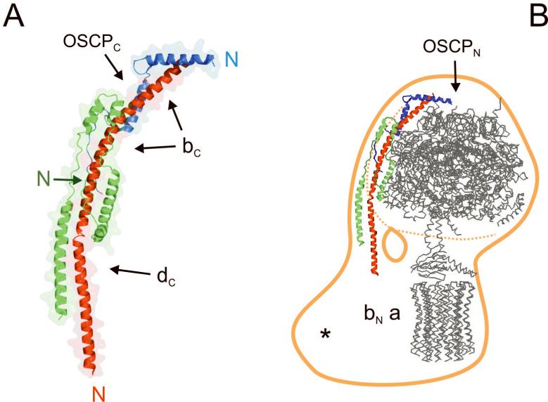
Structure and localization of the stator stalk of mitochondrial ATP synthase. (A) Structure of the b/d/F6 subcomplex [10]. Subunit b is depicted in red, subunit d in green, subunit F6 in blue. For each subunit, the N-terminus of the portion included in the crystallized subcomplex is marked. Black arrows indicate where some of the subunit fragments not included in the crystallized subcomplex might be accomodated. bC, C-terminus of subunit b; dC, C-terminus of subunit d; OSCPC, C-terminus of subunit OSCP (OSCP corresponds to the bacterial subunit δ in Fig. 1). (B) Localization of the stator stalk. The b/d/F6 subcomplex [10] (red/green/blue) was docked by eye to the F1c10subcomplex [11] (grey), using RasMol (www.bernstein-plus-sons.com), and both were modeled into a model of mitochondrial ATP synthase derived by cryo-electron microscopy (orange; after [12], with permission). The approximate location of subunit a, the N-terminus of b (“bN”), and the N-terminus of OSCP (“OSCPN”) is indicated. Mitochondrial ATP synthase contains a number of supernumerary proteins in the transmembrane region (marked with an asterisk) that are absent in the bacterial and chloroplast enzymes.
Similar articles
-
ATP synthase: subunit-subunit interactions in the stator stalk.Biochim Biophys Acta. 2006 Sep-Oct;1757(9-10):1162-70. doi: 10.1016/j.bbabio.2006.04.007. Epub 2006 Apr 19. Biochim Biophys Acta. 2006. PMID: 16730323 Free PMC article. Review.
-
Rows of ATP synthase dimers in native mitochondrial inner membranes.Biophys J. 2007 Oct 15;93(8):2870-6. doi: 10.1529/biophysj.107.109728. Epub 2007 Jun 8. Biophys J. 2007. PMID: 17557793 Free PMC article.
-
Assembly of human mitochondrial ATP synthase through two separate intermediates, F1-c-ring and b-e-g complex.FEBS Lett. 2015 Sep 14;589(19 Pt B):2707-12. doi: 10.1016/j.febslet.2015.08.006. Epub 2015 Aug 20. FEBS Lett. 2015. PMID: 26297831
-
Structural organization of mitochondrial ATP synthase.Biochim Biophys Acta. 2008 Jul-Aug;1777(7-8):592-8. doi: 10.1016/j.bbabio.2008.04.027. Epub 2008 Apr 27. Biochim Biophys Acta. 2008. PMID: 18485888
-
The peripheral stalk of the mitochondrial ATP synthase.Biochim Biophys Acta. 2006 May-Jun;1757(5-6):286-96. doi: 10.1016/j.bbabio.2006.01.001. Epub 2006 Jan 26. Biochim Biophys Acta. 2006. PMID: 16697972 Review.
Cited by
-
Transcriptome analysis of the bloom-forming dinoflagellate Prorocentrum donghaiense exposed to Ginkgo biloba leaf extract, with an emphasis on photosynthesis.Environ Sci Pollut Res Int. 2024 Mar;31(12):18579-18592. doi: 10.1007/s11356-024-32409-8. Epub 2024 Feb 13. Environ Sci Pollut Res Int. 2024. PMID: 38351353
-
Role of Charged Residues in the Catalytic Sites of Escherichia coli ATP Synthase.J Amino Acids. 2011;2011:785741. doi: 10.4061/2011/785741. Epub 2011 Jul 13. J Amino Acids. 2011. PMID: 22312470 Free PMC article.
-
Parasite powerhouse: A review of the Toxoplasma gondii mitochondrion.J Eukaryot Microbiol. 2022 Nov;69(6):e12906. doi: 10.1111/jeu.12906. Epub 2022 May 4. J Eukaryot Microbiol. 2022. PMID: 35315174 Free PMC article. Review.
-
Crystallization of the c14-rotor of the chloroplast ATP synthase reveals that it contains pigments.Biochim Biophys Acta. 2008 Jul-Aug;1777(7-8):605-12. doi: 10.1016/j.bbabio.2008.05.009. Epub 2008 May 19. Biochim Biophys Acta. 2008. PMID: 18515064 Free PMC article.
-
Evolutionary primacy of sodium bioenergetics.Biol Direct. 2008 Apr 1;3:13. doi: 10.1186/1745-6150-3-13. Biol Direct. 2008. PMID: 18380897 Free PMC article.
References
-
- Wilkens S. Rotary molecular motors. Adv. Protein Chem. 2005;71:345–382. - PubMed
-
- Weber J, Senior AE. ATP synthesis driven by proton transport in F1F0-ATP synthase. FEBS Lett. 2003;545:61–70. - PubMed
-
- Capaldi RA, Aggeler R. Mechanism of the F1F0-type ATP synthase, a biological rotary motor. Trends Biochem. Sci. 2002;27:154–160. - PubMed
-
- Boyer PD. A research journey with ATP synthase. J. Biol. Chem. 2002;277:39045–39061. - PubMed
-
- Carbajo RJ, et al. Solution structure of subunit F6 from the peripheral stalk region of ATP synthase from bovine heart mitochondria. J. Mol. Biol. 2004;342:593–603. - PubMed
Publication types
MeSH terms
Substances
Grants and funding
LinkOut - more resources
Full Text Sources
